Verlag für Medizin und Wirtschaft
Artikel
Bilder
Volltext
Mobile Version
A-
|
A
|
A+
Werbung
Abo
Bestellung Jahresabonnement (Print/elektronisch)
Bestellung kostenloses e-Journal-Abo
Mediadaten
Journale
News
Datenbanken
Suche Medizinische Publikationen
Suche Abbildungen und Grafiken
Medizinprodukte - Labortechnik
Antidotarium (Stand 2012)
Phytokodex (Stand 2012)
Endoskopie in Österreich (Stand 2010)
Kardiologie in Österreich (Stand 2006)
Osteologie in Österreich (Stand 2016)
Fortbildung
Bücher
Fachbücher
Patientenliteratur
Impressum
Offenlegung
Kontakt
Disclaimers & Copyright
Allgemeine Geschäftsbedingungen
Datenschutzerklärung
Sitemap
Figures and Graphics
Hoang-Xuan K et al.
Case Report: A 52-Year-Old HIV-Seropositive Patient with Hodgkin's Disease
European Association of NeuroOncology Magazine 2012; 2 (1)
: 45
PDF
Figures
Figure
Maximize View
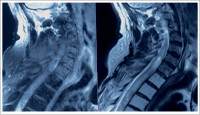
Thoracic Spinal Cord
Figure 1: Enlargement of the cervical and upper thoracic spinal cord with a hyperintense signal in a T2-weighted image from C3 to T7, with an intramedullary contrastenhancing lesion on C6.
Keywords:
MRI
,
spinal cord
,
Thorax
copyright © 2000–2025
Krause & Pachernegg GmbH
|
Sitemap
|
Datenschutz
|
Impressum