Brain - MRI
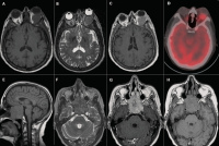
Figure 1a-h: MRI of the brain shows cranial plasmacytoma in a 61-year-old woman with MM IgA kappa who presented with left exophthalmos. (A) Axial T1-weighted
image shows an isointense mass in the left orbit. (B) Axial T2-weighted image shows the mass hypointense. (C) Axial T1-weighted gadolinium-enhanced image shows mild
enhancement of the mass. (D) Fused CT and FDG-PET images. (E, F, G, H) MRI of the brain shows a plasmacytoma of the clivus in a 57-year-old woman presenting with
diplopia and left-abducens palsy. The sagittal (E) and axial (H) T1-weighted images show an isointense mass arising from the clivus. (F) The axial T2-weighted image shows
a hypointense mass. (G) The axial T1-weighted gadolinium-enhanced image shows mild enhancement of the plasmacytoma.
Keywords:
brain,
MRI

