Krause und Pachernegg
Verlag für Medizin und Wirtschaft
Artikel   Bilder   Volltext

Mobile Version
A-  |   A  |   A+
Werbung
 
Figures and Graphics
Pace A, Villani V  
Extra-Cranial Metastases from Glioblastoma Multiforme

European Association of NeuroOncology Magazine 2012; 2 (2): 91-92

PDF    Figures   


Figure
 
Spine
Figure 1: Spine T1-weighted MRI image fat-sat showing a diffuse signal alteration in the vertebral bodies plus the presence of tissue at the lumbo-sacral level.


Keywords: lumbo-sacral levelMRIspine
 
 
Spine
Figure 2: Spine T1-weighted MRI image with gadolinium. The red arrow indicates the epidural tissue in the epidural space at the lumbosacral level.


Keywords: lumbo-sacral levelMRIspine
 
 
Spine
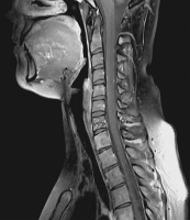
Figure 3: Cervical spine T1-weighted MRI image with gadolinium showing a mild leptomeningeal enhancement.


Keywords: leptomeningeal enhancementMRIspine
 
 
Skeletal involvement
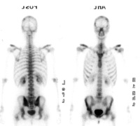
Figure 4: Radionuclide scan showing extensive skeletal involvement with diffuse vertebral and iliac bilateral localizations.


Keywords: radionuclide scanskeletal involvement


copyright © 2000–2025 Krause & Pachernegg GmbH | Sitemap | Datenschutz | Impressum