Krause und Pachernegg
Verlag für Medizin und Wirtschaft
Artikel   Bilder   Volltext

Mobile Version
A-  |   A  |   A+
Werbung
 
Figures and Graphics
Scherer M et al.  
Journey of Intraoperative Magnetic Resonance Imaging into Daily Use: A Review

European Association of NeuroOncology Magazine 2013; 3 (2): 61-67

PDF    Summary    Figures   


Figure
 
Seizures
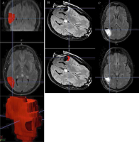
Figure 1a-c: 33-year-old male with seizures diagnosed with a WHO °II astrocytoma by stereotactic biopsy. Complete tumour resection was scheduled. (A) Preoperative definition of target tumour volume of 31.72 cm3 on T2 FLAIR images. (B) On the first iMRI, a 1.92-cm3 residual tumour is detected at the ventral aspect of the resection cavity on sharp FLAIR images. (C) Postoperative MRI shows no signs of tumour remnants. iMRI guidance led to a fully successful procedure with no added neurologic morbidity.


Keywords: iMRItumour


copyright © 2000–2025 Krause & Pachernegg GmbH | Sitemap | Datenschutz | Impressum