Verlag für Medizin und Wirtschaft
Artikel
Bilder
Volltext
Mobile Version
A-
|
A
|
A+
Werbung
Abo
Bestellung Jahresabonnement (Print/elektronisch)
Bestellung kostenloses e-Journal-Abo
Mediadaten
Journale
News
Datenbanken
Suche Medizinische Publikationen
Suche Abbildungen und Grafiken
Medizinprodukte - Labortechnik
Antidotarium (Stand 2012)
Phytokodex (Stand 2012)
Endoskopie in Österreich (Stand 2010)
Kardiologie in Österreich (Stand 2006)
Osteologie in Österreich (Stand 2016)
Fortbildung
Bücher
Fachbücher
Patientenliteratur
Impressum
Offenlegung
Kontakt
Disclaimers & Copyright
Allgemeine Geschäftsbedingungen
Datenschutzerklärung
Sitemap
Figures and Graphics
Millesi M et al.
Current Surgical Strategies in the Treatment of Intracranial Meningiomas
European Association of NeuroOncology Magazine 2013; 3 (3)
: 112-117
PDF
Summary
Figures
Figure
Maximize View
Intracranial Meningioma
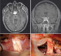
Figure 1: Tuberculum sellae meningioma found due to blurred vision operated via a right-sided subfrontal approach.
Keywords:
tuberculum sellae meningioma
Maximize View
Intracranial Meningioma
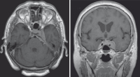
Figure 2: Left-sided cavernous-sinus meningioma found due to double vision treated by radiosurgery.
Keywords:
cavernous-sinus meningioma
Maximize View
Intracranial Meningioma
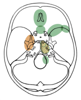
Figure 3: Current treatment strategies for skull base meningiomas. Green: surgical resection; brown: radiosurgery; green-brown: the best therapeutic option for petroclival meningiomas is still a matter of discussion.
Keywords:
scheme
,
skull base meningioma
,
treatment strategy
copyright © 2000–2025
Krause & Pachernegg GmbH
|
Sitemap
|
Datenschutz
|
Impressum