Krause und Pachernegg
Verlag für Medizin und Wirtschaft
Artikel   Bilder   Volltext

Mobile Version
A-  |   A  |   A+
Werbung
 
Figures and Graphics
Ayan F et al.  
Asymptomatic giant prolapsing right atrial myxoma: comparison of transthoracic and transesophageal echocardiography in pre-operative evaluation

Journal of Clinical and Basic Cardiology 2000; 3 (3): 197-198

PDF    Summary    Figures   


Figure
 
Atriales Myxom
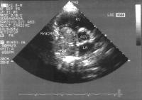
Figure 1: Transthoracic parasternal short-axis view showing a giant atrial myxoma in the almost totally occupied right atrium; RA, right atrium; RV, right ventricie; LA, left atrium; AOV, aorlic valve


Keywords: AtriumAtriumechocardiographyEchokardiographieMyxomMyxoma
 
 
Atriales Myxom
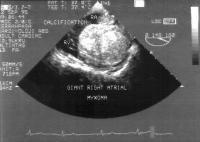
Figure 2: Transesophageal four-chamber view of the giant, lobulated and pedunculated myxoma showing calcifications in certain areas and attachment to the posterior wall; RA, right atrium; RV, right ventricle


Keywords: AtriumAtriumechocardiographyEchokardiographieMyxomMyxoma
 
 
Atriales Myxom
Figure 3A-B: Transesophageal four chamber view demonstrating a prolapsing giant right atrial myxoma; (A) Systole, (B) Diastole; RA, right atrium; RV, right ventricle; LA, left atrium; LV, left ventricle


Keywords: AtriumAtriumechocardiographyEchokardiographieMyxomMyxoma


copyright © 2000–2025 Krause & Pachernegg GmbH | Sitemap | Datenschutz | Impressum
 
Werbung