Krause und Pachernegg
Verlag für Medizin und Wirtschaft
Artikel   Bilder   Volltext

Mobile Version
A-  |   A  |   A+
Werbung
 
Figures and Graphics
Kozelj M et al.  
A calcified anterior mediastinal mass in a patient with aortic regurgitation

Journal of Clinical and Basic Cardiology 2000; 3 (3): 199-200

PDF    Summary    Figures   


Figure
 
Mediastinale Masse
Figure 1A-B: A postero-anterior chest radiograph shows a saccular, lobulated and well-delineated structure with a gently calcified eggshell-like contour in the heart silhouette on the right (white arrows) (A); a lateral chest radiograph shows this calcification in the lower retrosternal space (black arrows) (B).


Keywords: calcificationKalzifizierungMediastinumMediastinumRadiographRöntgenbild
 
 
Mediastinale Masse
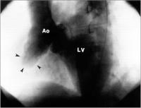
Figure 2: Contrast aortography in the left anterior oblique view revealed a huge aneurysm of the anterior wall of the ascending aorta (arrows), Ao = aorta, LV = left ventricle.


Keywords: aneurysmAneurysmaAngiographieangiographyAorta ascendensAorta ascendensMediastinumMediastinum
 
 
Mediastinale Masse
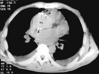
Figure 3: A CT scan of the chest revealed an aneurysm of the ascending aorta (Ao) with huge calcifications (arrows).


Keywords: aneurysmAneurysmaAorta ascendensAorta ascendensComputertomographiecomputertomographyMediastinumMediastinum


copyright © 2000–2025 Krause & Pachernegg GmbH | Sitemap | Datenschutz | Impressum
 
Werbung