Langendorff-Apparat
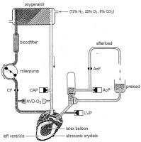
Figure 2: Modified Langendorff-Apparatus. The coronary perfusion circuit is shown on the left, and the systemic circuit is shown on the right. For details, see text.
Langendorff-Apparat - Herzfrequenz
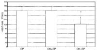
Figure 3: The heart rate (HR) was significantly reduced with DK. Mean +/- SD during pacing (EP), DK-AH 269 10-6 M (DK+EP) and without pacing (DK-EP), *p = 0.02
Langendorff-Apparat - linksventrikulärer Druck
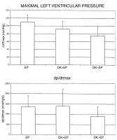
Figure 4: The peak left ventricular pressure (LVPmax) was reduced with DK (top); dP/dtmax was constant with DK+EP and was diminished without pacing (bottom). Mean +/- SD during pacing (EP), DK-AH 269 10-6 M (DK+EP) and without pacing (DK-EP)
Langendorff-Apparat - linksventrikulärer Druck
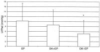
Figure 5: The end-diastolic left ventricular pressure (LVP ) was significantly decreased with DK after pacing was terminated. Mean +/- SD during pacing (EP), DK-AH 269 10-6 M (DK+EP) and without pacing (DK-EP), *p = 0.03
Langendorff-Apparat - koronarer Blutfluß
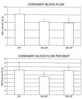
Figure 6: The global coronary blood flow (CBF) was reduced with DK+EP and remained constant after pacing was terminated (top) while the coronary blood flow per beat (CBF/beat) was increased in the last experimental step (bottom). Mean +/- SD during pacing (EP), DK-AH 269 10-6 M (DK+EP) and without pacing (DK-EP)
Langendorff-Apparat - myokardialer Sauerstoffverbrauch
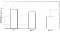
Figure 7: The myocardial oxygen consumption was decreased with DK+EP and furthermore with DK alone. Mean +/- SD during pacing (EP), DK-AH 269 10-6 M (DK+EP) and without pacing (DK-EP)