Krause und Pachernegg
Verlag für Medizin und Wirtschaft
Artikel   Bilder   Volltext

Mobile Version
A-  |   A  |   A+
Werbung
 
Figures and Graphics
Gasser S et al.  
Iatrogenic Coronary Fistula in Post Transplant Patients: Pathogenesis, Clinical Features and Therapy

Journal of Clinical and Basic Cardiology 2003; 6 (1-4): 19-21

PDF    Summary    Figures   


Figure
 
Koronarfistel
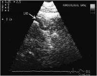
Figure 1: Transthoracic echocardiographic image of the right ventricle (RV) and the huge, dilated left coronary artery (left anterior descending branch; LAD) in a post transplant patient suffering from left coronary artery fistula into the right ventricle.


Keywords: coronary arteryechocardiographyEchokardiographieFistelfistulaKoronararterierechter Ventrikelright ventricle
 
 
Koronarfistel
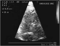
Figure 2: Post biopsy coronary fistula in a cardiac transplant patient. Colour flow imaging shows the drainage through the fistula into the right ventricle (white area above arrow which indicates insertion of the fistula). Transthoracal echocardiography constitutes an easily applicable means for the detection of post-biopsy coronary fistulae in transplant patients.


Keywords: BiopsiebiopsyechocardiographyEchokardiographieFistelfistularechter Ventrikelright ventricle
 
 
Koronarfistel
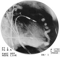
Figure 3: Dilated left anterior descending branch with small aneurysm and preinsertion stenosis draining into the right ventricle; the intensive flow through the fistula can be derived from a marked opacification of the right ventricle.


Keywords: AngiographieangiographyFistelfistularechter Ventrikelright ventricle


copyright © 2000–2025 Krause & Pachernegg GmbH | Sitemap | Datenschutz | Impressum
 
Werbung