Verlag für Medizin und Wirtschaft
Artikel
Bilder
Volltext
Mobile Version
A-
|
A
|
A+
Werbung
Abo
Bestellung Jahresabonnement (Print/elektronisch)
Bestellung kostenloses e-Journal-Abo
Mediadaten
Journale
News
Datenbanken
Suche Medizinische Publikationen
Suche Abbildungen und Grafiken
Medizinprodukte - Labortechnik
Antidotarium (Stand 2012)
Phytokodex (Stand 2012)
Endoskopie in Österreich (Stand 2010)
Kardiologie in Österreich (Stand 2006)
Osteologie in Österreich (Stand 2016)
Fortbildung
Bücher
Fachbücher
Patientenliteratur
Impressum
Offenlegung
Kontakt
Disclaimers & Copyright
Allgemeine Geschäftsbedingungen
Datenschutzerklärung
Sitemap
Abbildungen und Graphiken
Wewalka F et al.
Späte Perforation des proximalen Endes einer Pigtail-Prothese ins Duodenum
Journal für Gastroenterologische und Hepatologische Erkrankungen 2005; 3 (1)
: 19-21
Volltext (PDF)
Abbildungen
Abbildung
Vollbildansicht
Bulbus doudeni
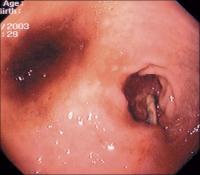
Abbildung 1: Bulbus duodeni mit perforierter proximaler Spitze der Gallengangsprothese.
Keywords:
Bulbus duodeni
,
Endoskopie
,
Gallengangprothese
,
Gastroenterologie
Vollbildansicht
Pigtail-Prothese - Papilla Vateri
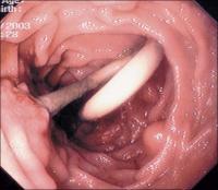
Abbildung 2: Tumoröse Papille mit liegender Pigtail-Prothese.
Keywords:
Endoskopie
,
Gastroenterologie
,
Papilla-Vateri
,
Pigtail-Prothese
Vollbildansicht
Doppel-Pigtail-Prothese - Gallengang
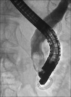
Abbildung 3: Doppel-Pigtail-Prothese im Gallengang; Perforation des proximalen Endes ins Duodenum
Keywords:
Duodenum
,
ERCP
,
Gastroenterologie
,
Pigtail-Prothese
copyright © 2000–2025
Krause & Pachernegg GmbH
|
Sitemap
|
Datenschutz
|
Impressum
Werbung