Krause und Pachernegg
Verlag für Medizin und Wirtschaft
Artikel   Bilder   Volltext

Mobile Version
A-  |   A  |   A+
Werbung
 
Figures and Graphics
Gasser S et al.  
Free Intracellular Magnesium Remains Uninfluenced by Changes of Extracellular Magnesium in Cardiac Guinea Pig Papillary Muscle

Journal of Clinical and Basic Cardiology 2005; 8 (1-4): 29-32

PDF    Summary    Figures   


Figure
 
Mg2+-ion-selective microelectrode
Figure 1: Mg2+-ion-selective microelectrode photographed by electrone microscopy. The ion-selective ligand can be seen in the tip of the microelectrode. The liquid junction potential indicating the individual Mg2+-concentration measured, arises at the surface between the reference solution and the ion-selective resin.


Keywords: ion-selectiveIonselektionSchemascheme
 
 
Mg2+
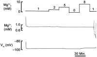
Figure 2: Changing extracellular concentrations of Mg2+ does not influence intracellular magnesium levels. One can see a typical experiment lasting approximately 3 hours. The signal stabilises some minutes after impalement. The electrode is equilibrated at the end of the experiment. Upon changing extracellullar Mg2+ a small depolarisation of the membrane potential can be seen without influencing the Mg2+-subtraction potential.


Keywords: diagramDiagrammMg++


copyright © 2000–2025 Krause & Pachernegg GmbH | Sitemap | Datenschutz | Impressum
 
Werbung