Krause und Pachernegg
Verlag für Medizin und Wirtschaft
Artikel   Bilder   Volltext

Mobile Version
A-  |   A  |   A+
Werbung
 
Figures and Graphics
Petzsch M, Reisinger EC  
Infective Endocarditis: Diagnostic and Therapeutic Issues - Should Transoesophagal Echocardiography be Performed in all Patients? - Position Pro

Journal of Clinical and Basic Cardiology 2001; 4 (2): 157-159

PDF    Summary    Figures   


Figure
 
Endokarditis - Diagnose - TEE
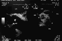
Figure 1: These abscesses were detected by TEE in a patient with IE of the aortic valve due to Staphylococcus lugdunensis. The infectious process extended from the aortic valve to the tissue between the aorta and the lateral border of the left atrium adjacent to the left atrial appendage. LA = left atrium; RA = right atrium; AV = aortic valve; MV = mitral valve; PA = pulmonary artery.


Keywords: AbscessechocardiographyEchokardiographieendocarditisEndokarditisStaphylococcus lugdunensisStaphylococcus lugdunensisTEETEE
 
 
Endokarditis - Diagnose - TEE
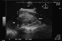
Figure 2: This abscess formed lateral and posterior of the aorta adjacent to the right atrium in a patient with IE due to Staphylococcus aureus. LA = left atrium; RA = right atrium; SVC = superior Vena cava.


Keywords: AbscessAortaAortaAtriumAtriumechocardiographyEchokardiographieendocarditisEndokarditisStaphylococcus aureusStaphylococcus aureus


copyright © 2000–2025 Krause & Pachernegg GmbH | Sitemap | Datenschutz | Impressum
 
Werbung