Krause und Pachernegg
Verlag für Medizin und Wirtschaft
Artikel   Bilder   Volltext

Mobile Version
A-  |   A  |   A+
Werbung
 
Abbildungen und Graphiken
Koulas S et al.  
Komplexe Rekonstruktion in der Gefäßchirurgie - Die tiefe Beinvene als alternatives Gefäßersatzmaterial

Zeitschrift für Gefäßmedizin 2010; 7 (3): 16-20

Volltext (PDF)    Summary    Praxisrelevanz    Abbildungen   


Abbildung
 
Plattenepithelkarzinom
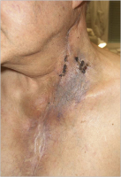
Abbildung 1: Patient 6 Wochen nach Resektion eines Plattenepithelkarzinoms und Neck Dissection mit Invasion in die Gefäßwand, welches zur gefäßchirurgischen Resektion und Rekonstruktion der Arteria carotis communis mit Lappendeckung belassen wurde.


Keywords: FotoGefäßmedizinPlattenepithelkarzinom
 
 
A. carotis communis
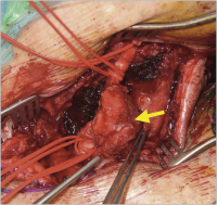
Abbildung 2: Freigelegte Carotis communis, proximal und peripher doppelt angeschlungen (gelber Pfeil: Plattenepithelkarzinom in die Karotiswand infiltrierend).


Keywords: Arteria carotis communisGefäßmedizin
 
 
Arteria carotis communis
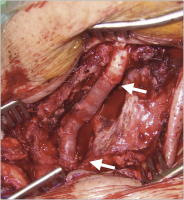
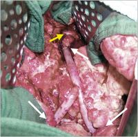
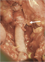
Abbildung 3: Nach Resektion der Carotis communis und des Tumors und Interponat mit tiefer Beinvene (weiße Pfeile: Anastomosen).


Keywords: Arteria carotis communisGefäßmedizin
 
 
Gefäßersatzmaterial
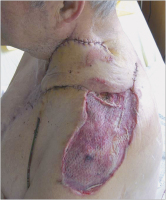
Abbildung 4: 5 Tage nach gefäßchirurgischer Rekonstruktion und plastischer Deckung


Keywords: FotoGefäßmedizin
 
 
Stenose
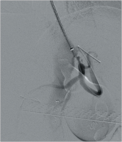
Abbildung 5: Hochgradige Stenose des Truncus coeliacus; Verschluss der Mesenterica superior und inferior (vor endovaskulärer Rekonstruktion).


Keywords: GefäßmedizinStenoseTruncus coeliacus
 
 
Bypass
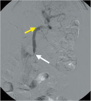
Abbildung 6: Bypass von der infrarenalen Aorta auf die Arteria lienalis. Hochgradige zentrale Abgangsstenose (weißer Pfeil) und Verschluss zur A. gastrica sinistra und hepatica communis (gelber Pfeil).


Keywords: Arteria lienalisBypassGefäßmedizin
 
 
Bypass
Abbildung 7: Aorto-mesenterica superior Vena-femoralis-Bypass. Aortale Anastomose (weißer Pfeil), Anastomose auf die A. mesenetrica superior (gelber Pfeil).


Keywords: BypassGefäßmedizinVena femoralis
 
 
Bypass
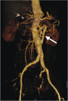
Abbildung 8: CT-Kontrolle 6 Monate postoperativ. Retrograde Füllung des Truncus coeliacus (Bypass: weißer Pfeil).


Keywords: BypassCTGefäßmedizin
 
 
Aortenaneurysma
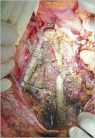
Abbildung 9: Zustand nach Aortenaneurysma und ischämischer Kolitis; nach Dickdarmresektion bestehender Protheseninfekt.


Keywords: AortenaneurysmaGefäßmedizin
 
 
Anastomose - Nierenvene
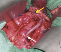
Abbildung 10: Nach Entfernung des Fremdmaterials und Rekonstruktion mit tiefer Beinvene. Anastomosen (weißer Pfeil), Nierenvene (gelber Pfeil).


Keywords: AnastomoseGefäßmedizinNierenvene
 
 
CT
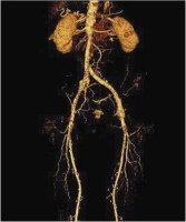
Abbildung 11: CT-Kontrolle 1 Jahr postoperativ.


Keywords: CTGefäßmedizin
 
 
Aneurysma
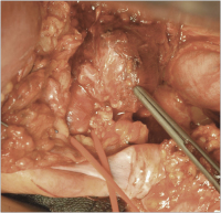
Abbildung 12: Aneurysma der Arteria iliaca communis bei Zustand nach Ileumpouch und lebensbedrohlicher Arrosionsblutung (Aneurysma an Pinzettenspitze; Iliaca externa angeschlungen).


Keywords: AneurysmaArteria iliaca communisGefäßmedizin
 
 
Rekonstruktion
Abbildung 13: Zustand nach Rekonstruktion mit tiefer Beinvene. Proximale Anastomose (weißer Pfeil).


Keywords: GefäßmedizinRekonstruktion


copyright © 2000–2025 Krause & Pachernegg GmbH | Sitemap | Datenschutz | Impressum
 
Werbung