Krause und Pachernegg
Verlag für Medizin und Wirtschaft
Artikel   Bilder   Volltext

Mobile Version
A-  |   A  |   A+
Werbung
 
Figures and Graphics
Siddiqui MA, Khan IA  
Transgastric and Transoesophageal Echocardiographic Views of Right Atrial Appendage

Journal of Clinical and Basic Cardiology 2001; 4 (4): 301

PDF    Summary    Figures   


Figure
 
Rechtsatrialer Appendix
Figure 1: Transgastric echocardiographic view of right-sided cardiac chambers in tangential plane of 153 degree. The right atrial appendage (A) is visualized in its longitudinal plane. RA = right atrium, RV = right ventricle, TV = tricuspid valve


Keywords: AppendixAtriumechocardiographyEchokardiographieright atrial appendage
 
 
Rechtsatrialer Appendix
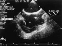
Figure 2: Transoesophageal echocardiographic view of both atria in longitudinal plane of 119 degree. The right atrial appendage (A) is visualized in its horizontal plane. IVC = inferior vena cava, LA = left atrium, RA = right atrium, RV = right ventricle, SVC = superior vena cava, TV = tricuspid valve, W = pacemaker wire


Keywords: AppendixAtriumechocardiographyEchokardiographieright atrial appendage
 
 
Rechtsatrialer Appendix
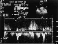
Figure 3: The pulsed Doppler evaluation of right atrial appendage through 154 degree transgastric view. The pulsed Doppler sample volume is shown in right upper corner of the figure.


Keywords: AppendixAtriumechocardiographyEchokardiographieright atrial appendage


copyright © 2000–2025 Krause & Pachernegg GmbH | Sitemap | Datenschutz | Impressum
 
Werbung