Neuhold U Echokardiographie aktuell: Gelungene Implantation eines Defibrillators mit Resynchronisationsfunktion, aber dem Patienten geht es schlechter ... Journal für Kardiologie - Austrian Journal of Cardiology 2008; 15 (9-10): 314-319 Volltext (PDF) Fallbeschreibung Übersicht
| ||||||||||||||||||
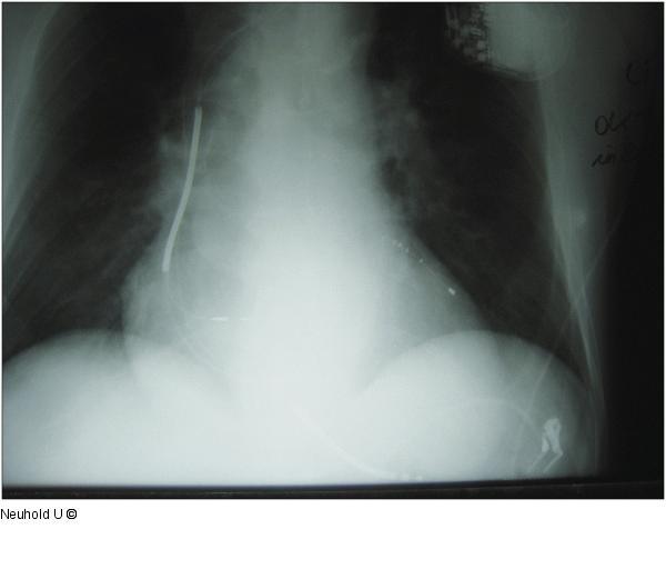
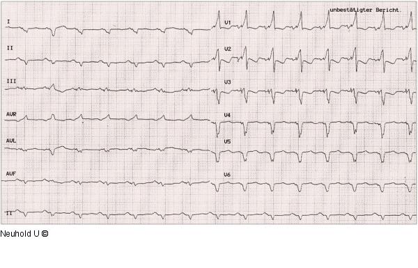
Abbildung 8a-b: c/p-Röntgen a) Rö c/p post-operativ; b) EKG mit biventrikulärem Pacing (typ. hohe R-Welle in V1) |
Abbildung 8a 
Abbildung 8b 
Abbildung 8a-b: c/p-Röntgen
a) Rö c/p post-operativ; b) EKG mit biventrikulärem Pacing (typ. hohe R-Welle in V1) |


