Peters S, Trümmel M, Koehler B Case report: Shorter-than-normal QT interval and provocable right precordial ST segment elevation in three patients with suspicious arrhythmogenic right ventricular cardiomyopathy Journal für Kardiologie - Austrian Journal of Cardiology 2011; 18 (9-10): 326-328 Volltext (PDF) Übersicht
| ||||||
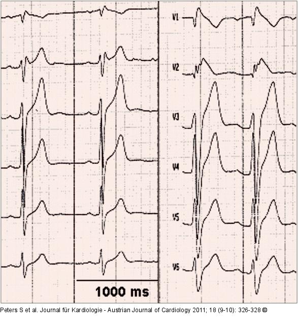
Abbildung 1: ECG leads Left: Standard precordial ECG leads V1–V6 (10 mV amplitude, 50 mm/s paper speed) of case no.1 with prolonged QRS duration in right precordial leads, small epsilon potential in V2 and saddle-back ST-segment elevation in V2. QTc interval 340 ms (NOTICE: horizontal line marking 1000 ms); right: precordial leads of case no. 1 after ajmaline administration with coved-type ST elevation in V1 and V2 and right bundle branch block configuration. |

Abbildung 1: ECG leads
Left: Standard precordial ECG leads V1–V6 (10 mV amplitude, 50 mm/s paper speed) of case no.1 with prolonged QRS duration in right precordial leads, small epsilon potential in V2 and saddle-back ST-segment elevation in V2. QTc interval 340 ms (NOTICE: horizontal line marking 1000 ms); right: precordial leads of case no. 1 after ajmaline administration with coved-type ST elevation in V1 and V2 and right bundle branch block configuration. |


