Azza MA, Ragab SH, Ismail NA, Awad MAM, Kandil ME Echocardiographic Assessment of Epicardial Adipose Tissue in Obese Children and Its Relation to Clinical Parameters of the Metabolic Syndrome Journal of Clinical and Basic Cardiology 2011; 14 (1-4): 7-11 PDF Summary Overview
| ||||||
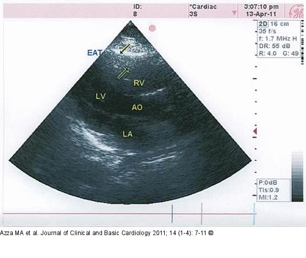
Figure/Graphic 1: EAT Transthoracic echocardiography showing a large area of epicardial adipose tissue (EAT) on the free wall of the right ventricle (arrows). |

Figure/Graphic 1: EAT
Transthoracic echocardiography showing a large area of epicardial adipose tissue (EAT) on the free wall of the right ventricle (arrows). |


