Montag M, Toth B, Strowitzki T Sperm Selection in ART Journal für Reproduktionsmedizin und Endokrinologie - Journal of Reproductive Medicine and Endocrinology 2012; 9 (6): 485-489 Volltext (PDF) Summary Übersicht
| ||||||
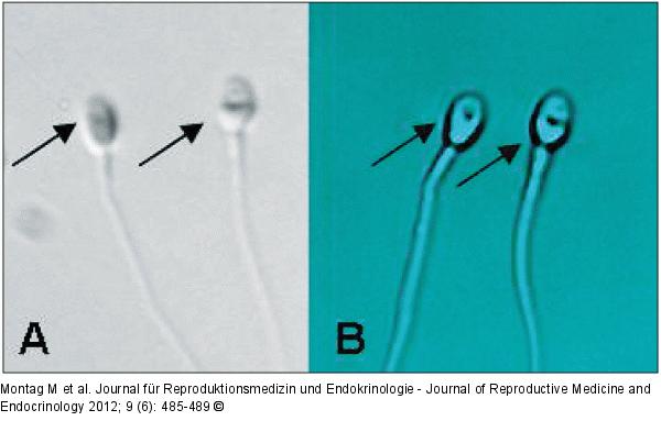
Abbildung 2a-b: Sperm Comparison of digital enhanced high magnification image versus conventional Hoffman modulation contrast. This figure shows a picture of two sperm taken with a standard Hofmann 40× objective (A) and a 63× objective with a high numerical aperture and using Cytoscreen (B) a digital image enhancement software (Octax, Bruckberg, Germany). The presence of a small vacuole in the head of the right sperm as visualized by digital enhanced high magnification can be seen in the standard Hofmann contrast picture as a irregularity in the sperm head. Reprinted from [23]. |

Abbildung 2a-b: Sperm
Comparison of digital enhanced high magnification image versus conventional Hoffman modulation contrast. This figure shows a picture of two sperm taken with a standard Hofmann 40× objective (A) and a 63× objective with a high numerical aperture and using Cytoscreen (B) a digital image enhancement software (Octax, Bruckberg, Germany). The presence of a small vacuole in the head of the right sperm as visualized by digital enhanced high magnification can be seen in the standard Hofmann contrast picture as a irregularity in the sperm head. Reprinted from [23]. |


