Asklund T, Henriksson R Radiotherapy and Meningioma European Association of NeuroOncology Magazine 2013; 3 (3): 122-127 PDF Summary Overview
| ||
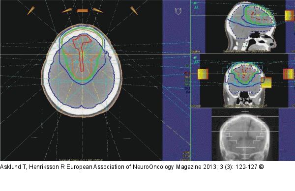
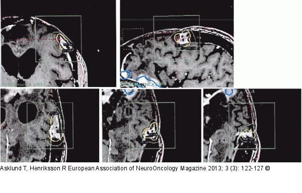
Figure/Graphic 1a-b: Radiotherapy - Meningioma Figures 1a and b depict the dose plan of 3-dimensional conformal radiotherapy and a dose plan of stereotactic radiosurgery (Gammaknife®), respectively, delivered to the patient described in the case report. In Figure 1b, the upper row represents, from left to right, a coronal and a sagittal section, while the lower row shows selected transaxial sections of the MR-based dose plan. |
Figure/Graphic 1a 
Figure/Graphic 1b 
Figure/Graphic 1a-b: Radiotherapy - Meningioma
Figures 1a and b depict the dose plan of 3-dimensional conformal radiotherapy and a dose plan of stereotactic radiosurgery (Gammaknife®), respectively, delivered to the patient described in the case report. In Figure 1b, the upper row represents, from left to right, a coronal and a sagittal section, while the lower row shows selected transaxial sections of the MR-based dose plan. |

