Schwabl C, Gaugg M, Beham-Schmid C, Celedin S, Liegl-Atzwanger B, Matschnig S, Raunik W, Weiss H, Neumann HJ Rheumatoide Arthritis und seltene Tumormanifestation – auf Umwegen zur Remission Tumorboard 2014; 3 (1): 5-9 Volltext (PDF) Übersicht
| ||||||||||||||
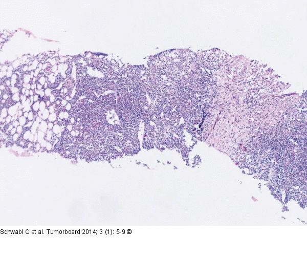
Abbildung 4a: Follikuläres Lymphom Biopsie (aus M. iliacus) mit Sklerosierung und dichter lymphatischer Infiltration. HE-Färbung, x 25. |

Abbildung 4a: Follikuläres Lymphom
Biopsie (aus M. iliacus) mit Sklerosierung und dichter lymphatischer Infiltration. HE-Färbung, x 25. |


