Grisold W, Grisold A Nerve Infiltration: Where, When, and How? An Introduction European Association of NeuroOncology Magazine 2014; 4 (2): 58-60 PDF Overview
| ||||
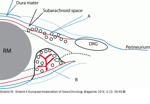
Figure/Graphic 1: Meningeal infiltration Meningeal infiltration. The dura mater continues as the perineurium. In general the tumor cells do not spread beyond this boundary centrifugally. (A) tumour spread in the subarachnoid space. (B) Lumps and nodules consist of tumour cells and have their own vascularisation. RM: spinal cord; DRG: dorsal root ganglion |

Figure/Graphic 1: Meningeal infiltration
Meningeal infiltration. The dura mater continues as the perineurium. In general the tumor cells do not spread beyond this boundary centrifugally. (A) tumour spread in the subarachnoid space. (B) Lumps and nodules consist of tumour cells and have their own vascularisation. RM: spinal cord; DRG: dorsal root ganglion |


