Baehring JM Lymphoma Nerve Infiltration European Association of NeuroOncology Magazine 2014; 4 (2): 61-64 PDF Summary Overview
| ||||
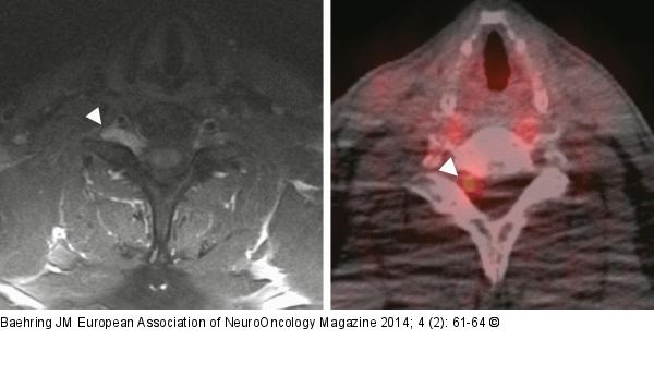
Figure/Graphic 2a-b: Primary neurolymphomatosis Magnetic resonance imaging shows a thickened and enhancing lower cervical nerve root in a patient with primary neurolymphomatosis ([a] T1-weighted sequence after administration of gadolinium). 18-Fluorodeoxyglucose positron emission tomography reveals increased tracer uptake within the affected nerve root ([b] arrow head). |

Figure/Graphic 2a-b: Primary neurolymphomatosis
Magnetic resonance imaging shows a thickened and enhancing lower cervical nerve root in a patient with primary neurolymphomatosis ([a] T1-weighted sequence after administration of gadolinium). 18-Fluorodeoxyglucose positron emission tomography reveals increased tracer uptake within the affected nerve root ([b] arrow head). |


