Bellmann B, Deppe I, Nagel P, Roser M, Muntean BG Case Report: Pacemaker Mimics Ventricular Extrasystoles? Journal für Kardiologie - Austrian Journal of Cardiology 2017; 24 (11-12): 286-287 Volltext (PDF) Übersicht
| ||||||
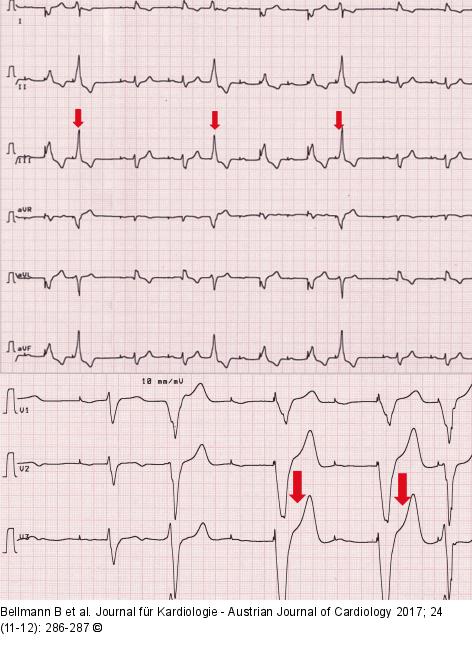
Abbildung 1: Ventricular premature beats Resting ECG with recordings of ventricular premature beats with left bundle branch block morphology. These are marked by the red arrow and show a partial pseudo-fusion. |

Abbildung 1: Ventricular premature beats
Resting ECG with recordings of ventricular premature beats with left bundle branch block morphology. These are marked by the red arrow and show a partial pseudo-fusion. |


