Weihs W, Schuchlenz H Echokardiographie aktuell: 3D-TEE bei PFO-Verschluss: Nützliches Tool oder aufwendiges Spielzeug? Journal für Kardiologie - Austrian Journal of Cardiology 2021; 28 (1-2): 40-41 Volltext (PDF) Übersicht
| ||||||||||
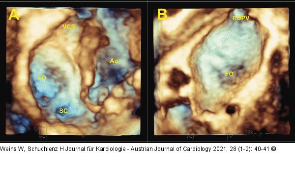
Abbildung 2: Echo Darstellungs des atrialen Septums von rechtsatrial (A) sowie nach Rotation um 180° von linksatrial (B) FO: Fossa ovalis, SC: Sinus coronarius, VCS: Vena cava superior, Ao: Aorta, RUPV: rechte obere Lungenvene. |

Abbildung 2: Echo
Darstellungs des atrialen Septums von rechtsatrial (A) sowie nach Rotation um 180° von linksatrial (B) FO: Fossa ovalis, SC: Sinus coronarius, VCS: Vena cava superior, Ao: Aorta, RUPV: rechte obere Lungenvene. |


