Valenta I, Upadhyaya A, Leucker T, Schindler TH Cardiac positron emission tomography in the identification and characterization of coronary artery disease and microvascular dysfunction // Kardiale PET zur Identifikation und Charakterisierung koronarer Herzkrankheiten und mikrovaskulärer Dysfunktion Journal für Kardiologie - Austrian Journal of Cardiology 2022; 29 (1-2): 34-40 Volltext (PDF) Summary Übersicht
| ||||
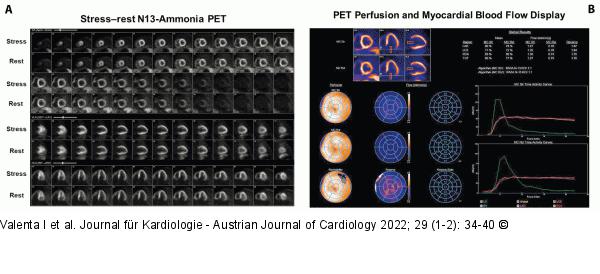
Abbildung 2: PET 13N-ammonia PET in the evaluation of CMD. Value of PET-determined hyperemic MBFs and MFR in the identification and characterization of CMD. A 65-year-old woman with recurrent chest pain and shortness of breath during daily activities. Cardiovascular risk profile with obesity (body mass index of 40 kg/m2 ), dyslipidemia, and hypertension. (A): Regadenoson-stress and rest 13N-ammonia PET/CT images in corresponding short-axis (top), vertical long-axis (middle), and horizontal long-axis (bottom) slices. On stress-images and rest perfusion images, there is homogenous radiotracer uptake of the left ventricle. Thus, no reversible radiotracer uptake to suggest regional ischemia and normal rest perfusion. Hemodynamically obstructive CAD lesions therefore can be widely ruled out. Gated peak-stress and rest PET images (not shown) demonstrated normal left ventricular size and wall motion associated with a left ventricular ejection fraction of 50% and 52%, respectively. (B): Display of quantitative MBF data unravel globally reduced hyperemic MBFs with only 1.29 mL/g/min (normal value > 1.80 mL/g/min) and decreased MFR with 1.71 (normal value > 2.0) to signify moderately severe CMD that may reflect a functional substrate for reported anginal symptoms. |

Abbildung 2: PET
13N-ammonia PET in the evaluation of CMD. Value of PET-determined hyperemic MBFs and MFR in the identification and characterization of CMD. A 65-year-old woman with recurrent chest pain and shortness of breath during daily activities. Cardiovascular risk profile with obesity (body mass index of 40 kg/m2 ), dyslipidemia, and hypertension. (A): Regadenoson-stress and rest 13N-ammonia PET/CT images in corresponding short-axis (top), vertical long-axis (middle), and horizontal long-axis (bottom) slices. On stress-images and rest perfusion images, there is homogenous radiotracer uptake of the left ventricle. Thus, no reversible radiotracer uptake to suggest regional ischemia and normal rest perfusion. Hemodynamically obstructive CAD lesions therefore can be widely ruled out. Gated peak-stress and rest PET images (not shown) demonstrated normal left ventricular size and wall motion associated with a left ventricular ejection fraction of 50% and 52%, respectively. (B): Display of quantitative MBF data unravel globally reduced hyperemic MBFs with only 1.29 mL/g/min (normal value > 1.80 mL/g/min) and decreased MFR with 1.71 (normal value > 2.0) to signify moderately severe CMD that may reflect a functional substrate for reported anginal symptoms. |


