Pranevicius R, Kanoun Schnur SS, Zirlik A, Toth GG, Harb S OCT-Corner: The importance of imaging in everyday percutaneous coronary intervention practice Journal für Kardiologie - Austrian Journal of Cardiology 2022; 29 (9-10): 266-269 Volltext (PDF) Übersicht
| ||||||||||||||||
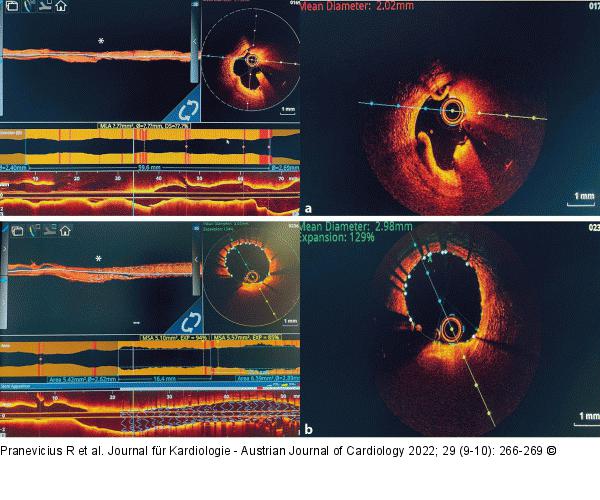
Abbildung 7: OCT OCT images before and after procedure of the same lesion. (a): Mean minimum diameter are of the tight stenosis before PCI; (b): Mean minimum diameter area after the PCI (*narrowest lesion of the LAD vessel). |

Abbildung 7: OCT
OCT images before and after procedure of the same lesion. (a): Mean minimum diameter are of the tight stenosis before PCI; (b): Mean minimum diameter area after the PCI (*narrowest lesion of the LAD vessel). |


