Krause und Pachernegg
Verlag für Medizin und Wirtschaft
Artikel   Bilder   Volltext

Mobile Version
A-  |   A  |   A+
Werbung
 
Bergmann M
Pleuraerguss – diagnostische Abklärung und therapeutische Möglichkeiten // Pleural fluid – diagnostic assessment and therapeutic options
Journal für Pneumologie 2024; 12 (2): 17-22

Volltext (PDF)    Summary    Übersicht   

Zum ersten Bild Abb. 1: Fibrinfäden Abb. 2: Septierter Erguss Abb. 3: Linsenförmiger Erguss Abb. 4a: Septierter Erguss Abb. 4b: Malignes Pleuramesotheliom Aktuelles Bild - Abb. 5: Epitheloides Mesotheliom Abb. 6: Thorakoskopische Talkumpleurodese Abb. 7: Abgeplattetes/invertiertes Zwerchfell
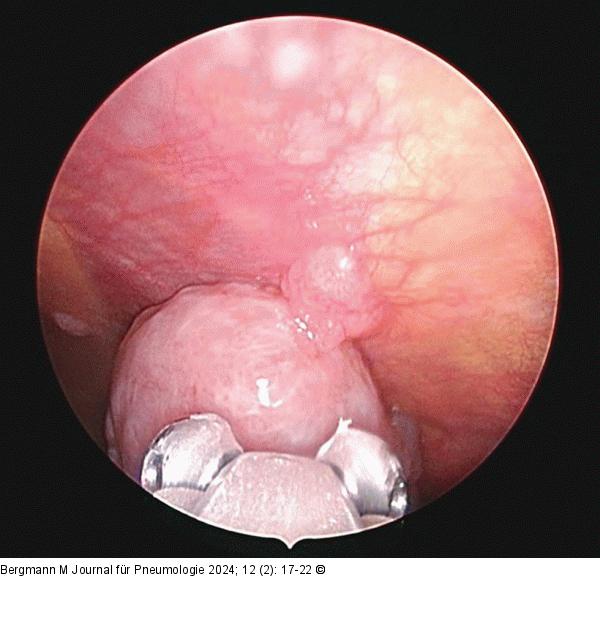
Abbildung 5: Epitheloides Mesotheliom
Epitheloides Mesotheliom (thorakoskopische Biopsie)
 
Epitheloides Mesotheliom
Vorheriges Bild Nächstes Bild   


Abbildung 5: Epitheloides Mesotheliom
Epitheloides Mesotheliom (thorakoskopische Biopsie)
 
copyright © 2000–2025 Krause & Pachernegg GmbH | Sitemap | Datenschutz | Impressum
 
Werbung