Nicolosi GL Infective Endocarditis: Diagnostic and Therapeutic Issues - Should Transesophageal Echocardiography be Performed in all Patients? - Position Con Journal of Clinical and Basic Cardiology 2001; 4 (2): 161-164 PDF Summary Overview
| ||||||||
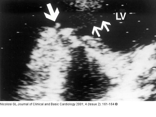
Figure/Graphic 3: Endokarditis - Diagnose - TEE Transthoracic modified 4 chamber apical view in a patient with a mitral mechanical prosthetic valve, where the disease was only evident on the ventricular side of the valve itself (large arrow), which was masking its presence by the transoesophageal approach. The two small arrows indicate a spontaneous echo contrast small diastolic jet through the stenotic prosthetic valve, due to ingrowing tissue (see text). LV = left ventricle |

Figure/Graphic 3: Endokarditis - Diagnose - TEE
Transthoracic modified 4 chamber apical view in a patient with a mitral mechanical prosthetic valve, where the disease was only evident on the ventricular side of the valve itself (large arrow), which was masking its presence by the transoesophageal approach. The two small arrows indicate a spontaneous echo contrast small diastolic jet through the stenotic prosthetic valve, due to ingrowing tissue (see text). LV = left ventricle |


