Kolb H-J, Böhm U, Mende M, Neugebauer A, Pfeiffer D, Rother T Assessment of the optimal atrioventricular delay in patients with dual chamber pacemakers using impedance cardiography and Doppler echocardiography Journal of Clinical and Basic Cardiology 1999; 2 (2): 237-240 PDF Summary Overview
| ||||||||
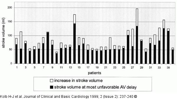
Figure/Graphic 3: AV-Intervall Increase in stroke volume (ml) following use of optimal atrioventricular delay measured by impedance cardiography at rest (VDD stimulation) |

Figure/Graphic 3: AV-Intervall
Increase in stroke volume (ml) following use of optimal atrioventricular delay measured by impedance cardiography at rest (VDD stimulation) |


