Gasser R, Horn S, Köppel H Discrimination by valinomycin K-selective surface microelectrodes of a sulphonylurea-sensitive and a distinct sulphonylurea-, barium-, TEA- and cinnamate-insensitive component of K-efflux from isolated pig coronary arteries during simulated ischaemia Journal of Clinical and Basic Cardiology 1998; 1 (1): 43-51 PDF Summary Overview
| ||||||||||||||||
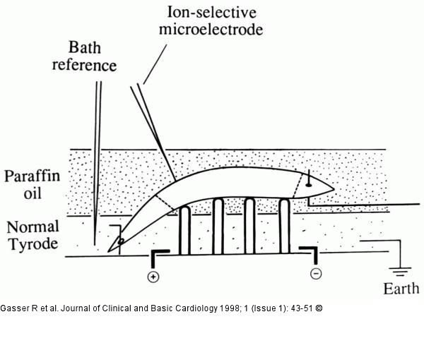
Figure/Graphic 1: K-Efflux in Koronararterien - Versuchsanordnung Experimental assembly used for simulating ischaemia. Fig. redrawn from Gasser & Vaughan-Jones (1990). Schematic diagram of perfusion chamber. Ion-selective microelectrode (in our experiments e.g. Na+, pH, K+- selective microelectrode) pressed gently on the surface of a pig coronary artery strip and recording specific ion concentration changes during ischaemia. Preparation is laid across four supports (micropins, 100 µm in diameter) and pinned on one end and attached to a force transducer at the other. Also shown are two electrodes for field Stimulation. The diagram illustrates an episode of simulated ischaemia, with the preparation immersed almost completely in a stationary pool of paraffin oil (heavy stippling) while perfusion of warm (37 °C) Tyrode (light stippling) continues at the base of the chamber. Dashed lines represent ligated areas as described in the Methods. |

Figure/Graphic 1: K-Efflux in Koronararterien - Versuchsanordnung
Experimental assembly used for simulating ischaemia. Fig. redrawn from Gasser & Vaughan-Jones (1990). Schematic diagram of perfusion chamber. Ion-selective microelectrode (in our experiments e.g. Na+, pH, K+- selective microelectrode) pressed gently on the surface of a pig coronary artery strip and recording specific ion concentration changes during ischaemia. Preparation is laid across four supports (micropins, 100 µm in diameter) and pinned on one end and attached to a force transducer at the other. Also shown are two electrodes for field Stimulation. The diagram illustrates an episode of simulated ischaemia, with the preparation immersed almost completely in a stationary pool of paraffin oil (heavy stippling) while perfusion of warm (37 °C) Tyrode (light stippling) continues at the base of the chamber. Dashed lines represent ligated areas as described in the Methods. |


