Hengstschläger M Stem Cells in Amniotic Fluid - What are the Next Steps to Do? Journal für Reproduktionsmedizin und Endokrinologie - Journal of Reproductive Medicine and Endocrinology 2005; 2 (4): 233-238 Volltext (PDF) Summary Übersicht
| ||
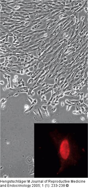
Abbildung 1: Oct-4 - Nucleus Detection of Oct-4 in the nucleus of a human amniotic fluid cell. A phase contrast picture of an amniotic fluid cell population grown under culture conditions used for routine prenatal diagnosis of genetic mutations is presented. For immunocytochemical analysis of cellular Oct-4 expression, cells were fixed and incubated with anti-Oct-4 antibody. Thereafter, cells were washed, incubated with a biotinylated secondary antibody, washed again, and incubated with ExtrAvidin-Cy3 conjugate. One amniotic fluid cell with the red Oct-4-specific signal in the nucleus is shown. |

Abbildung 1: Oct-4 - Nucleus
Detection of Oct-4 in the nucleus of a human amniotic fluid cell. A phase contrast picture of an amniotic fluid cell population grown under culture conditions used for routine prenatal diagnosis of genetic mutations is presented. For immunocytochemical analysis of cellular Oct-4 expression, cells were fixed and incubated with anti-Oct-4 antibody. Thereafter, cells were washed, incubated with a biotinylated secondary antibody, washed again, and incubated with ExtrAvidin-Cy3 conjugate. One amniotic fluid cell with the red Oct-4-specific signal in the nucleus is shown. |

