Michelmann HW, Schwartz P, Hinney B, Rath D Auf dem Weg der Samenzelle in die Eizelle entdeckt die Forschung immer noch neue Phänomene und Hindernisse Journal für Reproduktionsmedizin und Endokrinologie - Journal of Reproductive Medicine and Endocrinology 2006; 3 (1): 39-44 Volltext (PDF) Summary Übersicht
| ||||||||||||||||||
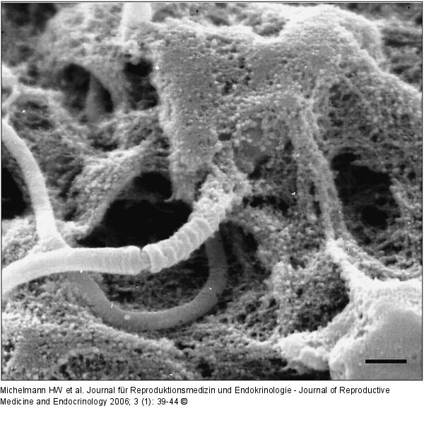
Abbildung 8: Spermatozoon - Zona pellucida Mit granulösem Zonamaterial überwucherter Kopf eines Spermatozoons in der Zona pellucida (Balken = 1 µm). |

Abbildung 8: Spermatozoon - Zona pellucida
Mit granulösem Zonamaterial überwucherter Kopf eines Spermatozoons in der Zona pellucida (Balken = 1 µm). |


