|
|
|---|
Unterbegriffe: AV-Angiom,
AVERT-Angiographie,
Abschluss-Angiographie,
Abschlussangiogramm,
Abschlussangiographie,
Akut-Koronarangiographie,
Akutangio,
Akutkoronarangiographie,
Amyloidangiopathie,
Angio-CT,
AngioDynamics,
AngioGuardsystem,
Angiocardiography,
Angiocentric,
Angiodysplasie,
Angiogene,
Angiogenese,
Angiografie,
Angiogram,
Angiogramm
|
1. Maaroufi A;
Andreka J;
Zirlik A;
Toth GG;
Harb S
OCT-Corner: It's all about the flow
Video 1
Journal für Kardiologie - Austrian Journal of Cardiology 2024; 31 (7-8): 183-185
Co-registration of OCT run in the LAD using Ultreon 2.0
software and Dragonfly Optis catheter. The video commences with
the proximal segment of the left anterior descending artery (LAD)
displaying simultaneous coronary angiogram on left and OCT imaging on right. Notably, the filling of the proximal LAD with contrast
appears suboptimal in the angiogram, concomitant with the OCT
images which vividly depict blood entering the vessel lumen. As the
video progresses distally, the OCT frames exhibit significant artifact
clearance from blood and better resolution of the vessel wall segments, concurrent with adequate contrast filling in the angiogram.
This is a nice example how artificial intelligence (AI) can prepare imaging data for quick and easy review by the interventional cardiologist:
In reality, blood is cleared from the lumen by antegrade dye injection
and the OCT-lens is moved from distal to proximal, so in this case it
reached the LM when blood already followed the contrast-bolus. AI
used the coregistered images and presents the video in antegrade
mode for more intuitive interpretation. Keep in mind, though, how
the pictures were created, to better understand the creation of artifacts. In this case, less experienced cardiologists may misinterprete
blood for thrombus if they only look at still frame OCT pictures. Co-registration of angio and OCT clearly shows the movement of blood.
“It is all about the flow!
Keywords: Kardiologie,
OCT-Corner
|
|
|
2. Mühlberger V;
Alber H;
Bonner G;
Brunner T;
Chatsakos T;
Christ G;
Delle Karth G;
Frick M;
Frömmel M;
Geppert A;
Grund M;
Haas M;
Hödl R;
Horcicka H;
Huber K;
Juhasz M;
Kaltenbach L;
Kaspar KD;
Mascherbauer J;
Neunteufl T
Herzkathetereingriffe in Österreich im Jahr 2020 (mit Audit 2021 in den Zentren der ANCALAR–Gruppe) // Cardiac Catheterization, Coronary Angiography (CA) and Percutaneous Coronary Intervention (PCI) in Austria during the Year 2020 (including AUDIT 2021)
PCI
Journal für Kardiologie - Austrian Journal of Cardiology 2022; 29 (3-4): 76-80
Anzahl der perkutanen koronaren Interventionen (PCI auf
der y-Achse) pro Million Einwohner in Österreich (AU) bis zum Jahr 2019
und hochgerechnet auf 100 % für das Jahr 2020 für AU, Schweiz (CH) bis
2018 [16] und Deutschland (D) bis 2019 [17, 18]. Schweden (SWE) ist abgeleitet aus „ Angio“; „PCI“; „PCI incl. Diagnostic“ [15] reproduziert für 2019
und 2020 aus dem SCAAR-Register. Keywords: Diagramm,
Kardiologie,
PCI
|
|
|
3. Mühlberger V;
Alber H;
Bonner G;
Brunner T;
Chatsakos T;
Christ G;
Delle Karth G;
Frick M;
Frömmel M;
Geppert A;
Grund M;
Haas M;
Hödl R;
Horcicka H;
Huber K;
Juhasz M;
Kaltenbach L;
Kaspar KD;
Mascherbauer J;
Neunteufl T
Herzkathetereingriffe in Österreich im Jahr 2020 (mit Audit 2021 in den Zentren der ANCALAR–Gruppe) // Cardiac Catheterization, Coronary Angiography (CA) and Percutaneous Coronary Intervention (PCI) in Austria during the Year 2020 (including AUDIT 2021)
CAG
Journal für Kardiologie - Austrian Journal of Cardiology 2022; 29 (3-4): 76-80
Anzahl der diagnostischen Koronar angiographien (CAG auf
der y-Achse) pro Million Einwohner in Österreich (AU) bis zum Jahr 2019
und hochgerechnet auf 100 % für das Jahr 2020 für AU, Schweiz (CH) bis
2018 [16] und Deutschland (D) bis 2019 [17, 18]. Schweden (SWE) ist abgeleitet aus „Angio“; „PCI“; „PCI incl. Diagnostic“ [15] reproduziert für 2019
und 2020 aus dem SCAAR-Register. Keywords: CAG,
Diagramm,
Kardiologie
|
|
|
4. Jacke C;
Hejazi S;
Marin G;
Görler U;
Arjumand J
Fallbericht: Thrombininjektion in die Art. obturatoria bei persistierender Beckenblutung
Persistierende Blutung
Journal für Kardiologie - Austrian Journal of Cardiology 2018; 25 (3-4): 82-83
Angio-CT zeigt die persistierende Blutung (weißer Pfeil) aus der linken Art. obturatoria. Keywords: Angio-CT,
Art. obturatoria,
Computertomographie,
Kardiologie,
persistierende Blutung
|
|
|
5. Holfeld J;
Tepeköylü C;
Schachner T
Bilder der Herzchirurgie: Hochalpiner Thoraxschmerz
CT-Angiographie
Journal für Kardiologie - Austrian Journal of Cardiology 2015; 22 (11-12): 292-294
In der CT- Angio wird eine akute Aortendissektion Stanford Typ A ersichtlich. Das Entry befindet sich im Aortenbogen, proximal sind bereits thrombosierte
Anteile des falschen Lumens sichtbar. Keywords: Angiographie,
CT
|
|
|
6. Pfabe FP
Das isolierte Iliakalaneurysma – Natürlicher Verlauf und aktueller Stand von Diagnostik und Therapie
A. iliaca communis
Zeitschrift für Gefäßmedizin 2014; 11 (3): 7-14
Endovaskuläre Ausschaltung eines Aneurysmas der A. iliaca communis
rechts (Typ I) mit fehlender distaler Landungszone. (a) Angiographischer Ausgangsbefund;
(b) Okkluderfreisetzung (Vascular Plug II, roter Pfeil); (c) erfolgreiche proximale
Embolisation der A. iliaca interna (roter Pfeil); (d) Abschlussbefund nach Embolisation
und Implantation eines linearen Grafts (Angio-CT in 3D-VRT). Keywords: 3D-VRT,
Angiographie,
Arteria iliaca communis,
Gefäßmedizin
|
|
|
7. Neugebauer P;
Jerabek P;
Kala P;
Bocek O;
Poloczek M;
Vytiska M;
Parenica J
Guiding Catheter-Induced Dissection with the Closure of the Last Patent Bypass Graft – A Case Report
Catheter-Induced Dissection
Journal für Kardiologie - Austrian Journal of Cardiology 2013; 20 (7-8): 222-225
Follow-up angio 4 months after PCI: 60% diffuse in-stent restenosis in tortuous middle part of LIMA, optimal result in LIMA-LAD anastomosis. Keywords: angiography,
Kardiologie,
LIMA-LAD anastomosis,
PCI,
percutaneous coronary intervention
|
|
|
8. Neugebauer P;
Jerabek P;
Kala P;
Bocek O;
Poloczek M;
Vytiska M;
Parenica J
Guiding Catheter-Induced Dissection with the Closure of the Last Patent Bypass Graft – A Case Report
Catheter-Induced Dissection
Journal für Kardiologie - Austrian Journal of Cardiology 2013; 20 (7-8): 222-225
Angio of RCA: diffuse 80–90% disease of proximal and middle part, small periphery. Keywords: angiography,
Kardiologie,
RCA,
right coronary artery
|
|
|
9. Neugebauer P;
Jerabek P;
Kala P;
Bocek O;
Poloczek M;
Vytiska M;
Parenica J
Guiding Catheter-Induced Dissection with the Closure of the Last Patent Bypass Graft – A Case Report
Catheter-Induced Dissection
Journal für Kardiologie - Austrian Journal of Cardiology 2013; 20 (7-8): 222-225
Angio of LCA: 80 % stenosis of distal left main stem (LMS), 90% stenosis of proximal LAD, closure of middle LAD and LCx. Keywords: angiography,
Kardiologie,
left main stem,
LMS,
Stenosis
|
|
|
10. Neugebauer P;
Jerabek P;
Kala P;
Bocek O;
Poloczek M;
Vytiska M;
Parenica J
Guiding Catheter-Induced Dissection with the Closure of the Last Patent Bypass Graft – A Case Report
Catheter-Induced Dissection
Journal für Kardiologie - Austrian Journal of Cardiology 2013; 20 (7-8): 222-225
Angio of LIMA: 90% stenosis in LIMA-LAD anastomosis. Keywords: angiography,
Kardiologie,
left internal mammary artery,
LIMA
|
|
|
11. Pfabe FP;
Hinc D
Fallbericht: Endovaskuläre Therapie eines retrograd perfundierten komplexen Iliaca-Aneurysmas nach aorto-bifemoraler Bypassanlage
Iliaca-Aneurysma
Zeitschrift für Gefäßmedizin 2013; 10 (1): 16-19
Visualisierung des minimalen Endoleaks Typ III (Pfeil) durch Angio-CT mit 3D-Rekonstruktion. Keywords: 3D-Rekonstruktion,
CT,
Endoleak,
Gefäßmedizin,
Iliaca-Aneurysma
|
|
|
12. Pfabe FP;
Hinc D
Fallbericht: Endovaskuläre Therapie eines retrograd perfundierten komplexen Iliaca-Aneurysmas nach aorto-bifemoraler Bypassanlage
Iliaca-Aneurysma
Zeitschrift für Gefäßmedizin 2013; 10 (1): 16-19
Ausgeprägte wandständige Thrombosierung des komplexen Aneurysmas in der CT- Angio in longitudinaler und sagittaler Ebene. Keywords: CT,
Gefäßmedizin,
Iliaca-Aneurysma,
Thrombose
|
|
|
13. Al-Nakkash A;
Zimmermann F
Fallbericht: Erfolgreiche Resektion eines zervikalen karotisnahen Schwannoms durch interdisziplinäres Management
CT
Zeitschrift für Gefäßmedizin 2010; 7 (3): 21-22
CT- Angio zeigt starke Tumorvaskularisation mit Verdrängung der Halsgefäße
nach ventral. Keywords: CT,
Gefäßmedizin,
Hals
|
|
|
14. Al-Nakkash A;
Rößler J;
Zimmermann F
Ungewähnlicher Verlauf einer Protheseninfektion mit erfolgreicher In-situ-Rekonstruktion und plastischer Deckung
Operationssitus
Zeitschrift für Gefäßmedizin 2008; 5 (2): 20-22
CT- Angio (Ausriss der aorto-bifemoralen Anastomosen) Keywords: CT-Angio,
Gefäßchirurgie,
Gefäßmedizin,
Phlebologie
|
|
|
15. Gruber A;
Bavinzski G;
Standhardt H;
Knosp E
Multimodale Therapie zerebraler Aneurysmen. Klinische, technische und angiographische Kriterien in der Entscheidungsfindung
Zerebrale Aneurysmen
Journal für Neurologie, Neurochirurgie und Psychiatrie 2007; 8 (3): 16-26
Indikationen und Kontraindikationen für die verfügbaren Techniken zur Behandlung zerebraler Aneurysmen. In dieser Matrix grün unterlegte Felder stellen allgemein gesprochen gängige bzw. zumindest mögliche Indikationen für die jeweilige
Therapieform dar, gelb unterlegte Felder problematische Konstellationen und
rot unterlegte Felder Kontraindikationen. Die Indikationen und Kontraindikationen
der "Balloon remodelling"-Technik decken sich weitgehend mit denen der endosakkulären
Coil-Embolisation. Da sich, anders als bei der Technik des "Balloon remodellings" und der Verwendung des nicht mehr verfügbaren "TriSpan Device", bei der stentgestützten Coil-Embolisation durch die erforderliche thrombozytenaggregationshemmende
Medikation spezielle Problemstellungen ergeben, wurde anstelle des Ãœberbegriffes
"Neck-bridging devices" in dieser Tabelle der Titel "Stent + Coil" gewählt. (Clip: mikrochirurgische Klippung; Coil: rekonstruktive endosakkuläre Embolisation; Stent + Coil: stentgestützte Coil-Embolisation; PAO: "Parent artery occlusion", d. h. therapeutischer Gefäßverschluß; PAO + EIAB: therapeutischer Gefäßverschluß unter Bypaßschutz; Flow Modif.: Flow-Modifikations-Techniken; "Small neck": enger
Aneurysmenhals; "Wide neck": breiter Aneurysmenhals; SAH +: rupturiertes Aneurysma;
SAH –: unrupturiertes Aneurysma; ICH: raumfordernde intrazerebrale Blutung
durch Aneurysmaruptur; Angio +: Aneurysma- und Gefäßmorphologie aus präoperativer
Bildgebung exakt bekannt; Angio –: suboptimale präoperative Bildgebung;
Morb +: erhebliche extrazerebrale Begleiterkrankungen mit erhöhtem Operationsund
Narkoserisiko; Morb –: keine wesentlichen Begleiterkrankungen; HH +: Hunt-
Hess-Stadium > 3; HH –: Hunt-Hess-Stadium ≤ 3; Ant. Circ.: Aneurysma der vorderen
Zirkulation; Post. Circ.: Aneurysma der hinteren Zirkulation; VSP +: posthämorrhagischer
Vasospasmus klinisch, angiographisch oder TCD sonographisch bzw. Behandlungszeitpunkt an Tag 4–12 nach Subarachnoidalblutung; VSP –: kein
Vasospasmus). Keywords: Aneurysma,
Behandlung,
Neurochirurgie,
Technik
|
|
|
16. Pürerfellner H;
Aichinger J;
Nesser H-J
Segmentale ostiale Katheterablation der Pulmonalvenen bei paroxysmalem Vorhofflimmern: Ergebnisse nach einjähriger Erfahrung
Pulmonalvenenangiographie
Journal für Kardiologie - Austrian Journal of Cardiology 2002; 9 (11): 497-505
PV- Angio: Selektive Darstellung der Einmündung der PV (RSPV) in das LA durch Kontrastmittelinjektion. Keywords: Angiographie,
Atrium cordis sinistrum,
Kardiologie,
Lassokatheter,
paroxysmales Vorhofflimmern,
Pulmonalvene,
PV
|
|
|
17. Kopp AF;
Claussen CD;
Heuschmid M;
Kuettner A;
Schroeder S
New Developments in Cardiac Imaging: The Role of MDCT
MDCT-Computertomographie
Journal of Clinical and Basic Cardiology 2001; 4 (4): 253-260
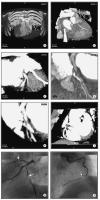
Male patient, age 55, with "situs inversus" and 2-vessel-disease, s/p PTCA of proximal RCA and prox. LAD, a 60 % lesion of distal RCA was left untreated. (a) Anterior view (volume rendering mode) of thorax with ventral thorax wall cut away depicts situs inversus. (b) RAO projection of RCX and prominent marginal branch in volume rendering mode. No highgrade lesion can be readily appreciated. (c) MIP of RCX clearly shows a
lesion (arrow). The degree of stenosis is estimated at 70 %. (d) RAO cranial view of proximal LAD in volume rendering mode. The high-grade lesion
was not clearly delineated in this view. (e) MIP-image clearly depicted the lesion of the proximal LAD. (f) MIP of RCA in "left posterior oblique"-projection. The arrow depicts the distal RCA lesion. The black arrow shows the area of the former dilatation, no restenosis present. (g) LAO cranial
view of LAD and RCX with conventional angio. The arrows depict the lesions in each of the vessels. (h) RAO view of RCA by conventional angiography.
Note the progress of the former 60 % lesion to a high-grade lesion and the absent restenosis of the
proximal RCA. Keywords: Arteria coronaria dextra,
Arteria coronaria dextra,
computer tomography,
Computertomographie,
MDCT,
MDCT,
Ramus circumflexus,
Ramus circumflexus,
Ramus interventricularis anterior,
Ramus interventricularis anterior,
Situs inversus,
Situs inversus,
two-vessel-disease,
Zweigefäßerkrankung
|
|
|
| |

 Suche Medizinische Publikationen
 Suche Abbildungen und Grafiken
 Medizinprodukte - Labortechnik
 Antidotarium (Stand 2012)
 Phytokodex (Stand 2012)
 Fachbücher
 Patientenliteratur