|
|
|---|
Unterbegriffe: Arteria arcuata,
Arteria basilaris,
Arteria brachialis,
Arteria carotis,
Arteria carotis communis,
Arteria carotis dextra,
Arteria carotis externa,
Arteria carotis interna,
Arteria cerebelli inferior posterior,
Arteria cerebelli interior posterior,
Arteria cerebelli superior,
Arteria cerebri anterior,
Arteria cerebri media,
Arteria cerebri media dextra,
Arteria cerebri posterior dextra,
Arteria cerebri posterior sinistra,
Arteria circumflexa,
Arteria communicans anterior,
Arteria coronaria,
Arteria coronaria cordis dextra
|
Oberbegriffe: Coro
Verwandte Begriffe: AVJR-„Sinus-coronarius“-Rhythmus,
Acute Coronary Syndrome,
Arteria coronaria,
Arteria coronaria cordis dextra,
Arteria coronaria dextra,
Arteria coronaria sinistra,
Corona,
Corona radiata,
Corona-radiata-Zellen,
Coronajahr,
Coronal,
Coronare,
Coronarschnitt,
Coronarsinus,
Coronary,
Coronary-CT,
Coronary-CT scan,
Coronavirus,
Sedacoron,
Sedacoron-Unverträglichkeit
|
1. Gangl C;
Delle Karth G
OCT-Corner: In-Stent-Restenose nach CTO
RCA - ISR - DEB
Journal für Kardiologie - Austrian Journal of Cardiology 2014; 21 (3-4): 98-100
RCA angiographisch nach Wiedereröffnung der In-Stent-Restenose mittels Drug-eluting Ballon Keywords: Angiographie,
Arteria coronaria dextra
|
|
|
2. Gangl C;
Delle Karth G
OCT-Corner: In-Stent-Restenose nach CTO
OCT- RCA - Stent
Journal für Kardiologie - Austrian Journal of Cardiology 2014; 21 (3-4): 98-100
OCT der beiden RCA-Stents post CTO-Wiedereröffnung Keywords: Arteria coronaria dextra,
OCT
|
|
|
|
|
|
4. Gangl C;
Delle Karth G
OCT-Corner: In-Stent-Restenose nach CTO
CTO - RCA
Journal für Kardiologie - Austrian Journal of Cardiology 2014; 21 (3-4): 98-100
RCA-CTO in Doppelinjektion mit teilweiser kollateraler Versorgung
aus dem linken Koronarsystem Keywords: Angiographie,
Arteria coronaria dextra
|
|
|
|
|
|
|
|
|
7. Weihs W;
Schuchlenz H;
Prein K
Echokardiographie aktuell: Kardiale Beteiligung bei Systemerkrankungen - Amyloidose
Koronararterie
Journal für Kardiologie - Austrian Journal of Cardiology 2010; 17 (7-8): 313-315
Rechte Koronararterie mit 2 hochgradigen Stenosen vor (linkes Bild) und nach der Stentimplantation (rechtes Bild). Keywords: Arteria coronaria dextra,
Echokardiographie,
Kardiologie
|
|
|
8. Schmid J;
Auer J
Aus dem Herzkatheterlabor: Seltene Differentialdiagnose eines akuten Koronarsyndroms bei einer älteren kaukasischen Patientin
Koronaraterie
Journal für Kardiologie - Austrian Journal of Cardiology 2010; 17 (11-12): 426-429
Rechte Koronararterie in LAO-Projektion. Keywords: Arteria coronaria dextra,
Kardiologie
|
|
|
9. Saurer G;
Weihs W
Fallbericht: Transient Left Ventricular Apical Ballooning Syndrome (Tako-Tsubo-Kardiomyopathie) - ein Fallbericht
Angiogramm - Koronararterie
Journal für Kardiologie - Austrian Journal of Cardiology 2006; 13 (11-12): 383-385
Unauffälliges Angiogramm der rechten Koronararterie. Keywords: Angiographie,
Arteria coronaria dextra,
Kardiologie
|
|
|
10. Auer J;
Eber B;
Maurer E
Akutes Koronarsyndrom bei Stenose des Hauptstammes der linken Herzkranzarterie
Akutes Koronarsyndrom - Stenose der linken Herzkranzarterie
Journal für Kardiologie - Austrian Journal of Cardiology 2004; 11 (4): 169-171
Progression in der proximalen rechten Herzkranzarterie mit einer nunmehr höchstgradigen Stenose Keywords: akutes Koronarsyndrom,
Angiographie,
Arteria coronaria dextra,
Kardiologie,
Stenose
|
|
|
11. Auer J;
Eber B;
Maurer E
Akutes Koronarsyndrom bei Stenose des Hauptstammes der linken Herzkranzarterie
Akutes Koronarsyndrom - Stenose der linken Herzkranzarterie
Journal für Kardiologie - Austrian Journal of Cardiology 2004; 11 (4): 169-171
50%ige Stenosierung in der RCA proximal Keywords: Angiographie,
Arteria coronaria dextra,
Kardiologie,
Stenose
|
|
|
12. Auer J
Aus dem Herzkatheterlabor: Transientes linksventrikuläres "Apical Ballooning" als Differentialdiagnose zum akuten Myokardinfarkt
Apical Ballooning
Journal für Kardiologie - Austrian Journal of Cardiology 2004; 11 (12): 522
Die Koronarangiographie zeigt keinen Hinweise für Stenosen an den großen epikardialen Gefäßen (links: rechte Koronararterie in LAO-Projektion; rechts: linke Koronararterie in LAO-Projektion) Keywords: Arteria coronaria dextra,
Arteria coronaria sinistra,
Kardiologie,
Koronarangiographie,
LAO-Projektion
|
|
|
13. Gyöngyösi M;
Fröhlich G;
Garamvölgyi R;
Glogar HD;
Heinisch B;
Hevesi A;
Losert U;
Matiasek J;
Pavo N;
Petrasi Z;
Repa I;
Scherzer TM;
Shirazi M;
Sperker W;
Strehblow C;
Windberger U;
Yahya N
Intrakoronare Stents: Der Weg von der Fabrik bis in die menschlichen Koronararterien - Präklinische Evaluierung der Koronarstents
Arteria coronaria - Hausschwein
Journal für Kardiologie - Austrian Journal of Cardiology 2004; 11 (12): 515-521
Linke Koronararterien (links) (LAD, LCx) und rechte Koronararterie (rechts) beim Hausschwein; AP-Aufnahme → Stent (2,75 mm/18 mm) in der LAD, Stent (4,0 mm/18 mm) in der LCx Keywords: AP-Aufnahme,
Arteria coronaria dextra,
Arteria coronaria sinistra,
Kardiologie,
Stent
|
|
|
14. Grebe O;
Hombach V
Neueste Entwicklungen in der bildgebenden kardialen Diagnostik
MR- Koronaranomalie - Perfusionsdefekt
Journal für Kardiologie - Austrian Journal of Cardiology 2003; 10 (9): 369-374
Koronaranomalie mit Abgang der rechten Kranzarterie aus dem linken Sinus valsalvae mit Verlauf zwischen Aorta und Pulmonalishauptstamm (links). Durch die Kompression kommt es zu einem unter Belastung verstärkten Perfusionsdefekt im Versorgungsbereich (Pfeile) Keywords: Arteria coronaria dextra,
Kardiologie,
Koronaranomalie,
Magnetresonanztomographie,
Perfusionsdefekt,
Sinus valsalvae
|
|
|
15. Globits S;
Kaiser B;
Mayr H;
Mittendorfer M;
Salomonowitz E
Koronargefäßdiagnostik mittels Magnetresonanztomographie
Kardiale Magnetresonanztomographie
Journal für Kardiologie - Austrian Journal of Cardiology 2003; 10 (1-2): 50-55
Patient mit kollateralem Verschluß der RCA. a) konventionelles Röntgenangiogramm der RCA; b) konventionelles Röntgenangiogramm der LCA mit Kollateralen zur RCA; c) 3D-bTFE über das Verschlußloch hinaus (Pfeil) Keywords: Arteria coronaria dextra,
Arteria coronaria sinistra,
Herz,
Kardiologie,
Magnetresonanztomogramm,
Magnetresonanztomographie,
Röntgenangiogramm,
Röntgenangiographie
|
|
|
16. Globits S;
Kaiser B;
Mayr H;
Mittendorfer M;
Salomonowitz E
Koronargefäßdiagnostik mittels Magnetresonanztomographie
Kardiale Magnetresonanztomographie
Journal für Kardiologie - Austrian Journal of Cardiology 2003; 10 (1-2): 50-55
Koronargefäßanomalie mit Abgang der LCX aus der RCA. M2D-TSE (black blood) links, 3D-bTFE in Atemanhaltetechnik (bright blood) rechts Keywords: Arteria coronaria dextra,
Herz,
Kardiologie,
Magnetresonanztomogramm,
Magnetresonanztomographie,
Ramus circumflexus
|
|
|
17. Globits S;
Kaiser B;
Mayr H;
Mittendorfer M;
Salomonowitz E
Koronargefäßdiagnostik mittels Magnetresonanztomographie
Kardiale Magnetresonanztomographie
Journal für Kardiologie - Austrian Journal of Cardiology 2003; 10 (1-2): 50-55
3D-bTFE + Navigatortechnik bei einem gesunden Probanden: oben links LAD gewinkelt axial; oben rechts: LAD gewinkelt sagittal; unten links: RCA gewinkelt koronal; unten rechts: LCX gewinkelt koronal Keywords: Arteria coronaria dextra,
Herz,
Kardiologie,
Magnetresonanztomogramm,
Magnetresonanztomographie,
Ramus circumflexus,
Ramus interventricularis anterior
|
|
|
|
|
|
19. Globits S;
Kaiser B;
Mayr H;
Mittendorfer M;
Salomonowitz E
Koronargefäßdiagnostik mittels Magnetresonanztomographie
Kardiale Magnetresonanztomographie
Journal für Kardiologie - Austrian Journal of Cardiology 2003; 10 (1-2): 50-55
Scout 2: bTFE + Navigatortechnik axial zur Identifikation der Koronargefäßabgänge (weißer Pfeil = RCA, schwarzer Pfeile = LAD) Keywords: Arteria coronaria dextra,
Herz,
Kardiologie,
Magnetresonanztomogramm,
Magnetresonanztomographie,
Navigatortechnik,
Ramus interventricularis anterior
|
|
|
|
|
|
21. Kaiser B;
Globits S;
Mayr H;
Mittendorfer M;
Salomonowitz E
Myokardiale First-Pass-Perfusionsdiagnostik mittels Magnetresonanztomographie
Kardiale Magnetresonanztomographie
Journal für Kardiologie - Austrian Journal of Cardiology 2003; 10 (1-2): 26-31
Männlicher Patient, 59 Jahre, St. p. Hinterwandinfarkt (5 Wochen alt), kollateralisierter Verschluß der rechten Koronararterie (RCA). a: CINE-Studie (Balanced-TFE-SENSE), Kontraktilitätsanalyse, kurze Achse, mittventrikulär, enddiastolisch; b: CINE-Studie (Balanced-TFE-SENSE), Kontraktilitätsanalyse, kurze Achse, mittventrikulär, endsystolisch, akinetisches Segment diaphragmal; c: First-Pass-Perfusionsstudie (SR-T1-TFE-SENSE), mittventrikuläre Schicht in kurzer Achse, subendokardialer Perfusionsdefekt diaphragmal; d: Vitalitätsstudie (3D-IR-TFE-SENSE), mittventrikuläre Schicht in kurzer Achse, 15 min nach i.v. KM-Applikation, signifikantes Late Enhancement diaphragmal (> 50 % transmyokardial, Avitalität/Narbe); e: Röntgen-Angiographie, RCA, proximal verschlossen; f: Röntgen-Angiographie, LAD mit Gefäßkollateralen zur RCA Keywords: Arteria coronaria dextra,
Herz,
Kardiologie,
Magnetresonanztomogramm,
Magnetresonanztomographie,
Röntgenangiogramm,
Röntgenangiographie,
Thorax
|
|
|
|
|
|
23. Auer J;
Eber B
Die Bedeutung der Kaliumkanalöffner für die Therapie der Angina pectoris
Koronarangiographie - Stenose - Stent
Journal für Kardiologie - Austrian Journal of Cardiology 2003; 10 (Supplementum C): 11-15
Links: Selektive Koronarangiographie mit Darstellung zweier Stenosen in der rechten Herzkranzarterie; Rechts: Selektive Koronarangiographie - Kontrollangiogramm nach PTCA (perkutane transluminale Koronarangioplastie) und Stentimplantation indie Stenosen in der rechten Herzkranzarterie Keywords: Angiogramm,
Arteria coronaria dextra,
Herzkranzarterie,
Kardiologie,
Koronarangiographie,
Stenose,
Stent
|
|
|
|
|
|
25. Hofmann R
Fallbericht: Abciximab verhindert STEMI
ST-Hebungs-Myokardinfarkt
Journal für Kardiologie - Austrian Journal of Cardiology 2002; 9 (12): 570-572
Angiogramm der rechten Koronararterie nach 4 Stunden: subtotale Stenose (Kreis), rechts nach Intervention mit einem Koronarstent. Keywords: Angiographie,
Arteria coronaria dextra,
Kardiologie,
Stenose,
Stent
|
|
|
26. Hofmann R
Fallbericht: Mobiler Thrombus in der RCA bei NSTEMI
Mobiler Thrombus der RCA bei NSTEMI
Journal für Kardiologie - Austrian Journal of Cardiology 2002; 9 (11): 510-511
Gleicher Koronargefäßabschnitt nach Stentimplantation: Füllungsdefekt nicht mehr nachweisbar. Keywords: Angiographie,
Arteria coronaria dextra,
Fallbericht,
Kardiologie,
Stent
|
|
|
27. Hofmann R
Fallbericht: Mobiler Thrombus in der RCA bei NSTEMI
Mobiler Thrombus in der RCA bei NSTEMI
Journal für Kardiologie - Austrian Journal of Cardiology 2002; 9 (11): 510-511
Angiogramm der rechten Koronararterie: diffuse nichtstenosierende sklerotische Veränderungen; Kontrastmittelübertragungsdefekt im mittleren Drittel (Kreis). Keywords: Angiographie,
Arteria coronaria dextra,
Fallbericht,
Kardiologie,
Sklerose,
Thrombus
|
|
|
28. Kopp AF;
Claussen CD;
Heuschmid M;
Kuettner A;
Schroeder S
New Developments in Cardiac Imaging: The Role of MDCT
MDCT-Computertomographie
Journal of Clinical and Basic Cardiology 2001; 4 (4): 253-260
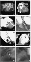
Male patient, age 55, with "situs inversus" and 2-vessel-disease, s/p PTCA of proximal RCA and prox. LAD, a 60 % lesion of distal RCA was left untreated. (a) Anterior view (volume rendering mode) of thorax with ventral thorax wall cut away depicts situs inversus. (b) RAO projection of RCX and prominent marginal branch in volume rendering mode. No highgrade lesion can be readily appreciated. (c) MIP of RCX clearly shows a
lesion (arrow). The degree of stenosis is estimated at 70 %. (d) RAO cranial view of proximal LAD in volume rendering mode. The high-grade lesion
was not clearly delineated in this view. (e) MIP-image clearly depicted the lesion of the proximal LAD. (f) MIP of RCA in "left posterior oblique"-projection. The arrow depicts the distal RCA lesion. The black arrow shows the area of the former dilatation, no restenosis present. (g) LAO cranial
view of LAD and RCX with conventional angio. The arrows depict the lesions in each of the vessels. (h) RAO view of RCA by conventional angiography.
Note the progress of the former 60 % lesion to a high-grade lesion and the absent restenosis of the
proximal RCA. Keywords: Arteria coronaria dextra,
Arteria coronaria dextra,
computer tomography,
Computertomographie,
MDCT,
MDCT,
Ramus circumflexus,
Ramus circumflexus,
Ramus interventricularis anterior,
Ramus interventricularis anterior,
Situs inversus,
Situs inversus,
two-vessel-disease,
Zweigefäßerkrankung
|
|
|
29. Kopp AF;
Claussen CD;
Heuschmid M;
Kuettner A;
Schroeder S
New Developments in Cardiac Imaging: The Role of MDCT
MDCT-Computertomographie
Journal of Clinical and Basic Cardiology 2001; 4 (4): 253-260
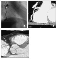
Male patient, age 62, known 2-vessel-disease, s/p PTCA of proximal LAD. Patient presented with recurrent chest–pain and angina. (a) RAO-projection of conventional angiogram reveals high grade stenosis of descending RCA. (b) LAO projection of RCA by MDCT (collimation 4 x 1 mm, pitch 1.5, 120 cc Imeron(R) 400) depicts the proximal lesion as well as wall changes throughout the vessel. (c) Axial CT image of the mid-lesion region: the degree of stenosis is estimated to 80 % (arrow). Keywords: Arteria coronaria dextra,
Arteria coronaria dextra,
computer tomography,
Computertomographie,
MDCT,
MDCT,
Stenose,
Stenosis,
two-vessel-disease,
Zweigefäßerkrankung
|
|
|
30. Kopp AF;
Claussen CD;
Heuschmid M;
Kuettner A;
Schroeder S
New Developments in Cardiac Imaging: The Role of MDCT
MDCT-Computertomographie
Journal of Clinical and Basic Cardiology 2001; 4 (4): 253-260
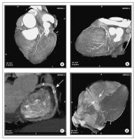
MDCT angiography (collimation 4 x 1 mm, pitch 1.5, 120 cc Imeron(R) 400). (a) Anterior view of left coronary artery with LAD in volume rendering technique. (b) Lateral view of left coronary artery with LAD and circumflex branch. (c) Maximum intensity projection of right coronary artery with calcified plaques (arrow). (d) Diaphragmatic surface with posterolateral and interventricular
branches of RCA (arrows). Keywords: Angiographie,
angiography,
Arteria circumflexa,
Arteria circumflexa,
Arteria coronaria dextra,
Arteria coronaria dextra,
computer tomography,
Computertomographie,
MDCT,
MDCT,
Ramus interventricularis anterior,
Ramus interventricularis anterior
|
|
|
| |

 Suche Medizinische Publikationen
 Suche Abbildungen und Grafiken
 Medizinprodukte - Labortechnik
 Antidotarium (Stand 2012)
 Phytokodex (Stand 2012)
 Fachbücher
 Patientenliteratur