| |
| Alle Journale wurden nach Diffusion durchsucht |
Treffer 1 - 13 von 13 |
|
|
|---|
Oberbegriffe: Diffus,
Fusion,
Sion
Unterbegriffe: Anisotrope Diffusion,
Diffusion-Perfusion-Impairment,
Diffusiongewichtung,
Diffusions-MRT,
Diffusionsgewichtete,
Diffusionskoeffizient,
Diffusionsstörung,
Diffusionstensors,
apparent diffusion coefficient,
diffusion weighted imaging,
diffusionsgewichtete MRT-Sequenzen,
periventrikuläre Diffusionsstörung
Verwandte Begriffe: AMI-Perfusion,
AV-Knotendepression,
Ablationsläsionen,
Accession,
Adhesion,
Adhäsion,
Adhäsions-Rezeptoren,
Adhäsionsbildung,
Aggression,
Aggressionsverhalten,
Ammoniak-Positronenemissionstomographie,
Ammoniak-Positronenemmissionstomographie,
Androgensuppression,
Aortenläsion,
Aquaporinexpression,
Arrosion,
Arrosionsblutung,
Arzneimittelkommission,
BG-Läsion,
Ballonokklusion
|
1. Unrath A;
Müller HP;
Kassubek J
Voxelbasierte MR-Morphometrie und Diffusionstensorbildgebung: Grundlagen und Anwendungen in der klinischen Neurologie
FA-Maps
Journal für Neurologie, Neurochirurgie und Psychiatrie 2011; 12 (3): 272-279
Fraktionale Anisotropie- (FA-) Maps eines gesunden Probanden, 36
Jahre, Hintergrund ist eine Aufnahme ohne Gradientengewichtung, Schwellenwert
für die FA-Darstellung ist 0,2. Farbkodierung: rot: größter Eigenvektor in Rechts-Links-
Richtung; blau: inferior-superior; grün: posterior-anterior. Keywords: Anisotrope Diffusion,
Neurologie
|
|
|
2. Unrath A;
Müller HP;
Kassubek J
Voxelbasierte MR-Morphometrie und Diffusionstensorbildgebung: Grundlagen und Anwendungen in der klinischen Neurologie
Anisotrope Diffusion
Journal für Neurologie, Neurochirurgie und Psychiatrie 2011; 12 (3): 272-279
Anisotrope Diffusion
von 1H entlang der Axone in einem
Voxel erlaubt die Berechnung eines
Diffusionstensors für dieses Voxel.
Die enthaltene Information kann als
Parametrisierung in „fractional
anisotropy (FA) maps“ oder als Fiber
Tracking verwendet werden. Keywords: Anisotrope Diffusion,
Neurologie
|
|
|
3. Bucek R;
Berzaczy D;
Popovic M;
Puchner S;
Reiter M
Zerebrale arterielle Verschlusskrankheit - CTA, MRA, MRT, Perfusions- und Diffusions-MR: Brauche ich das alles?
Stenose
Zeitschrift für Gefäßmedizin 2008; 5 (3): 10-14
Prä- und postinterventionelles Angiogramm eines
Patienten mit hochgradiger Stenose der A. carotis interna mit den korrespondierenden TTP-Maps
("time-to-peak") einer MR-Perfusionsstudie.
Die dunklen Areale zeigen präinterventionell eine deutliche Minderperfusion des rechten Mediastromgebietes bei symmetrischer
Perfusionssituation postinterventionell. Keywords: Angiographie,
Arteria carotis interna,
Gefäßmedizin,
Stenose
|
|
|
4. Bucek R;
Berzaczy D;
Popovic M;
Puchner S;
Reiter M
Zerebrale arterielle Verschlusskrankheit - CTA, MRA, MRT, Perfusions- und Diffusions-MR: Brauche ich das alles?
Plaque
Zeitschrift für Gefäßmedizin 2008; 5 (3): 10-14
B-Flow und Farbdopplersonographie einer echoarmen Plaque mit tiefem Ulcus Keywords: Farbdoppler,
Gefäßmedizin,
Plaque,
Sonographie
|
|
|
5. Bucek R;
Berzaczy D;
Popovic M;
Puchner S;
Reiter M
Zerebrale arterielle Verschlusskrankheit - CTA, MRA, MRT, Perfusions- und Diffusions-MR: Brauche ich das alles?
Plaque
Zeitschrift für Gefäßmedizin 2008; 5 (3): 10-14
Asymptomatische Plaque einer 70-jährigen Patientin – GSM 106; histologisch wurden ausgedehnte verkalkte und fibröse Anteile festgestellt. Keywords: CTA,
Gefäßmedizin,
Plaque
|
|
|
6. Bucek R;
Berzaczy D;
Popovic M;
Puchner S;
Reiter M
Zerebrale arterielle Verschlusskrankheit - CTA, MRA, MRT, Perfusions- und Diffusions-MR: Brauche ich das alles?
Plaque
Zeitschrift für Gefäßmedizin 2008; 5 (3): 10-14
Asymptomatische Plaque eines 82-jährigen Patienten – GSM 16; die histologische Auswertung ergab eine Fibroatheromplaque ohne Einblutung oder Ulzeration. Keywords: CTA,
Gefäßmedizin,
Plaque
|
|
|
7. Bucek R;
Berzaczy D;
Popovic M;
Puchner S;
Reiter M
Zerebrale arterielle Verschlusskrankheit - CTA, MRA, MRT, Perfusions- und Diffusions-MR: Brauche ich das alles?
Plaque
Zeitschrift für Gefäßmedizin 2008; 5 (3): 10-14
Symptomatische (Zustand nach brachiofazial betonter TIA) Plaque einer 78-jährigen Frau – GSM 9; die histologische Auswertung ergab eine eingeblutete
Atheromplaque. Keywords: CTA,
Gefäßmedizin,
Plaque
|
|
|
8. Schenk P;
Müller C
Das Hepatopulmonale Syndrom
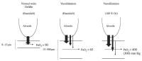
Diffusion - Perfusion - Impairment
Journal für Gastroenterologische und Hepatologische Erkrankungen 2004; 2 (3): 38-46
" Diffusion-Perfusion-Impairment": Schematische Darstellung des Pathomechanismus für die Hypoxämie beim HPS. O2-Moleküle
von den angrenzenden Alveolen können nicht in das Zentrum der extrem dilatierten Kapillaren diffundieren, um das Hämoglobin in den Erythrozyten zu oxygenieren. Dies kann durch das Einatmen von 100 % O2 über
eine Gesichtsmaske überwunden werden. Keywords: Alveole,
Hepatologie,
Hypoxämie,
Schema
|
|
|
9. Geser F;
Wenning GK
Klinik und Therapie der Multisystematrophie
MSA - Diffusion weighted imaging
Journal für Neurologie, Neurochirurgie und Psychiatrie 2004; 5 (2): 56-62
DWI ( Diffusion weighted imaging) bei a) der Parkinson-Erkrankung und b) der MSA-P. Blau, grün, rot und schwarz repräsentieren abnehmende
regionale ADC-(apparent diffusion coefficient-)Werte; es zeigt sich ein deutlich ausgeprägter Unterschied in den putaminalen ADC-Werten zwischen MSA-P (b) und PD (a). Keywords: apparent diffusion coefficient,
diffusion weighted imaging,
DWI,
Morbus Parkinson,
MSA-P,
Neurologie
|
|
|
10. Tentschert S;
Kreuzer S;
Lalouschek W;
Lang W;
Nasel Ch
Dissektion der Arteria carotis als Ursache ischämischer zerebrovaskulärer Ereignisse - Fallberichte und Diskussion
Gehirn - Perfusion
Journal für Neurologie, Neurochirurgie und Psychiatrie 2003; 4 (1): 13-20
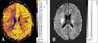
A) Perfusions-MRT: Es zeigt sich eine deutliche hämodynamische Belastung im Stromgebiet der Arteriae cerebri anterior et media dextra (rote und blaue Zonen rechts-telenzephal). B) Diffusions-MRT (b1000i-Bild): Innerhalb der deutlich hämodynamisch belasteten Areale ist ein Infarkt nachweisbar (weißes Areal cella media rechts) Keywords: Arteria cerebri anterior,
Arteria cerebri media,
Diffusion,
Gehirn,
Magnetresonanztomographie,
Neurologie,
Perfusion
|
|
|
11. Sabbah HN;
Goldstein S;
Sharov VG
Remodeling of the cardiac interstitium in the progression of heart failure
Herzinsuffizienz - Progression
Journal of Clinical and Basic Cardiology 1999; 2 (1): 113-116
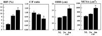
Bar graph (mean +/- SEM) depicting changes in volume fraction of reactive interstitial fibrosis (RIF), capillary density (C/F Ratio), oxygen diffusion distance (ODD) and average myocyte cross-sectional area (MCSA) in LV myocardium of normal dogs (NL), dogs with heart failure (CHF) sacrificed at 2 weeks (2w) and 4 months (4m) after the last embolization. *= p < 0.05 2w vs. 4m Keywords: Diagramm,
heart failure,
Herzinsuffizienz,
Progression,
Progression
|
|
|
12. Di Napoli P;
Barsotti A;
Contegiacomo G;
Di Iorio P;
Di Muzio M;
Giuliani P;
Maggi A;
Statile D;
Taccardi AA
Effects of A1 adenosine receptor antagonism against ischaemia-reperfusion damage and coronary microcirculation in isolated working rat hearts
Adenosin und koronare Mikrozirkulation
Journal of Clinical and Basic Cardiology 1999; 2 (1): 99-104
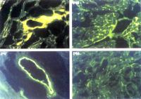
Histological samples (200x, fluorescent light) showing the FITC-albumin diffusion. A: control heart submitted to 20 min global ischaemia, B) effect of 100 nm DPCPX: a significant reduction of perivascular (PV, left) and perimyocytic (PM, right) fluorescence is detected. Keywords: Adenosin,
adenosine,
Histologisches Präparat,
histology,
ischaemia,
Ischämie,
microcirculation,
Mikrozirkulation
|
|
|
13. Di Napoli P;
Barsotti A;
Contegiacomo G;
Di Crecchio A;
Di Muzio M;
Maggi A;
Taccardi AA;
Tiloca P
Ischaemic preconditioning of rat myocardium: effects on postischaemic coronary endothelium hyperpermeability and microcirculatory damage
Preconditioning - koronare Mikrozirkulation
Journal of Clinical and Basic Cardiology 1998; 1 (1): 37-42
Histological samples (200X, fluorescent light) showing the FITC-Albumin diffusion in perimyocytic space (left) and perivascular space (right). Top: control. Middle: preconditioning. Bottom: preconditioning + hypertonic reperfusion. In control group fluorescence is strongly diffused around myocytes and perivascular space. After ischaemic preconditioning, albumin extravasation is limited; adding hypertonic reperfusion FlTC-albumin diffusion is detected in the only endothelial surface of blood vessels (bottom, on the right) Keywords: FITC-albumin,
FITC-albumin,
Histologisches Präparat,
histology,
ischaemic preconditioning,
ischämisches Preconditioning,
microcirculation,
Mikrozirkulation
|
|
|
| |
|

 Suche Medizinische Publikationen
 Suche Abbildungen und Grafiken
 Medizinprodukte - Labortechnik
 Antidotarium (Stand 2012)
 Phytokodex (Stand 2012)
 Fachbücher
 Patientenliteratur