| |
| Alle Journale wurden nach Endometrial durchsucht |
Treffer 1 - 10 von 10 |
|
|
|---|
Oberbegriffe: Endo,
Trial
Unterbegriffe: Subendometriale,
endometrial cyst,
endometrial lesion,
subendometriale Fibrose
Verwandte Begriffe: Aortenklappenendokarditis,
Atrial,
Atrial Natriuretic Peptide,
Atrial-hochseptal,
BHAT-Trial,
Ballonendoskopie,
Chromoendoskopie,
Crescendo-Decrescendo-artiger,
Darmendometriose,
Descendorektostomie,
Deszendostomie,
Endo1,
EndoAnchor,
EndoAnchor-Implantation,
EndoAnchor™-System,
EndoRotor®,
EndoV,
Endobag,
Endocrine,
Endocrinically
|
1. Götte M;
Kiesel L
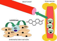
Endocrine Aspects of Endometrial Stem Cell Function in Reproductive-Age Women
Effects of estrogen on adult stem cell recruitment
Journal für Reproduktionsmedizin und Endokrinologie - Journal of Reproductive Medicine and Endocrinology 2013; 10 (Sonderheft 1): 120-125
Effects of estrogen on adult stem cell recruitment. Estrogen stimulates cytokine secretion by ER+ cells
withing the endometrial stem cell niche, leading to an activation of ER- endometrial stem cells (lower left panel). In addition, estrogen modulates the release of bone-marrow-derived CD133+ endothelial progenitor cells into the circulation right panel. See text for details. Keywords: cytokine,
Estrogen,
stem cell
|
|
|
2. Schweppe KW;
Rabe T;
Langhardt M;
Woziwodzki J;
Petraglia F;
Kiesel L
Endometriosis – Pathogenesis, Diagnosis, and Therapeutic Options for Clinical and Ambulatory Care
Adenomyosis uteri interna
Journal für Reproduktionsmedizin und Endokrinologie - Journal of Reproductive Medicine and Endocrinology 2013; 10 (Sonderheft 1): 102-119
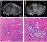
Adenomyosis uteri interna. (a): In the sonographic cross-section, the inhomogeneous appearance of the myometrium can be seen with a poorly delineated area of adenomyosis (unlike myomas); (b): Doppler sonography shows the increased perfusion in the area of adenomyosis (US images: Elsässer, Heidelberg); (c): The endometrial focus visible within the uterine muscles (HE, 12,5 ×); (d): With a higher magnification, the glands and the cytogenic stroma can be clearly discerned (HE, 40 ×) Keywords: adenomyosis,
uteri interna
|
|
|
3. Schweppe KW;
Rabe T;
Langhardt M;
Woziwodzki J;
Petraglia F;
Kiesel L
Endometriosis – Pathogenesis, Diagnosis, and Therapeutic Options for Clinical and Ambulatory Care
Endometrial cyst
Journal für Reproduktionsmedizin und Endokrinologie - Journal of Reproductive Medicine and Endocrinology 2013; 10 (Sonderheft 1): 102-119
Endometrial cyst in the area of the ovary. (a): Left-sided endometrial cyst of the ovary with adhesions
and concomitant reactive inflammation; (b): Ultrasound view of an endometrial cyst: homogeneous, low internal echo (bleeding) and thickened cyst wall; cap-shaped residual ovary on right picture margin (Elsässer, Heidelberg); (c): Extensive, endocrinically inactive endometrial cyst of the ovary with bleeding (HE, 25,5 ×); (d): Florid, endocrinically active endometrial cyst of the ovary within the hyperplastic cyst wall (HE, 40 ×). Keywords: endometrial cyst,
ovary
|
|
|
4. Schweppe KW;
Rabe T;
Langhardt M;
Woziwodzki J;
Petraglia F;
Kiesel L
Endometriosis – Pathogenesis, Diagnosis, and Therapeutic Options for Clinical and Ambulatory Care
Vicious circle in the endometrial focus
Journal für Reproduktionsmedizin und Endokrinologie - Journal of Reproductive Medicine and Endocrinology 2013; 10 (Sonderheft 1): 102-119
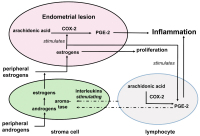
Vicious circle in the endometrial focus maintains the proliferation and inflammatory reaction: (1) local estrogen is produced by aromatase activation; (2) estrogens stimulate prostaglandin synthesis via the activation of the COX-2 enzyme; (3) Prostaglandin E-2 again stimulates aromatase. Whether the aromatase is activated in the endometrial lesion itself or in the surrounding zissue (fat, peritoneum) is controversial. Keywords: endometrial lesion,
metabolism,
Prostaglandin
|
|
|
5. Schweppe KW;
Rabe T;
Langhardt M;
Woziwodzki J;
Petraglia F;
Kiesel L
Endometriosis – Pathogenesis, Diagnosis, and Therapeutic Options for Clinical and Ambulatory Care
Estrogen metabolism in endometrial lesions
Journal für Reproduktionsmedizin und Endokrinologie - Journal of Reproductive Medicine and Endocrinology 2013; 10 (Sonderheft 1): 102-119
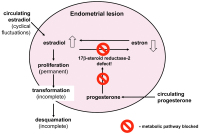
Estrogen metabolism in endometrial lesions: Defective 17-beta-steroid-dehydrogenase type 2. This means that the bioactive estradiol cannot be converted into the less active estron. In the normal endometrium, progesterone activates this 17-beta-HSD Type 2 and has an antiproliferative effect; this mechanism is disrupted in endometrial lesions (so-called progesterone block). Keywords: endometrial lesion,
estrogen metabolism
|
|
|
6. Schweppe KW;
Rabe T;
Langhardt M;
Woziwodzki J;
Petraglia F;
Kiesel L
Endometriosis – Pathogenesis, Diagnosis, and Therapeutic Options for Clinical and Ambulatory Care
Endocrinically active endometriosis
Journal für Reproduktionsmedizin und Endokrinologie - Journal of Reproductive Medicine and Endocrinology 2013; 10 (Sonderheft 1): 102-119
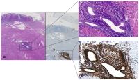
Microscopic image of endocrinically active endometriosis of the appendix (E = intramural endometrial focus; M = mucous membrane of the appendix). (a): highly differentiated, proliferative endometrial foci (HE stain, 200 ×); (b): highly positive estrogen receptors (ERp, brown stain, 200 l). Keywords: appendicitis,
endometriosis,
salpingitis
|
|
|
7. Schweppe KW;
Rabe T;
Langhardt M;
Woziwodzki J;
Petraglia F;
Kiesel L
Endometriosis – Pathogenesis, Diagnosis, and Therapeutic Options for Clinical and Ambulatory Care
Endoscopic appearance of endometriosis
Journal für Reproduktionsmedizin und Endokrinologie - Journal of Reproductive Medicine and Endocrinology 2013; 10 (Sonderheft 1): 102-119
Endoscopic appearance of endometriosis. (a): fresh peritoneal endometrial lesion on the pelvic peritoneum with distinctly visible vessels (Rimbach/Saarlouis); (b): Older, nodular peritoneal endometriosis in the area of the lig. sacrouterinum (Rimbach/Saarlouis); (c): Endometriosis in the area of the tube wall with peritubal adhesions (Rimbach/Saarlouis); (d): Conglomerate of appendix and tube in endometriosis: The infundibulum and the inflamed appendix stick together, mixed with vesicular, partially bloody endometriosis (Endo). Keywords: endometriosis,
macroscopic manifestation
|
|
|
8. Schweppe KW;
Rabe T;
Langhardt M;
Woziwodzki J;
Petraglia F;
Kiesel L
Endometriosis – Pathogenesis, Diagnosis, and Therapeutic Options for Clinical and Ambulatory Care
Adenocarcinoma
Journal für Reproduktionsmedizin und Endokrinologie - Journal of Reproductive Medicine and Endocrinology 2013; 10 (Sonderheft 1): 102-119
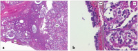
(a): Small-cell adenocarcinoma (CC) in the cyst wall of ovarian endometriosis [EM] (HE stain, magnification 12.5 ×); (b): Highly prismatic epithelium lining the endometrial cyst of the ovary [EM]; next to it in the same ovary, a clear-cell adenomcarcinoma [CC] (HE stain, magnification 200 ×) Keywords: adenocarcinoma,
clear-cell,
ovary,
small-cell
|
|
|
9. Fitzgerald JS;
Poehlmann TG;
Suman P;
Gupta SK;
Schleussner E;
Markert UR
Signal Transducer and Activator of Transcription 3 (STAT3) and Trophoblast Invasion
Implantation window
Journal für Reproduktionsmedizin und Endokrinologie - Journal of Reproductive Medicine and Endocrinology 2007; 4 (6): 322-330
Plate 1 depicts gradual apposition of blastocyst to endometrium during the onset of the implantation
window, delineated here by putative biomarkers
for endometrial receptivity: integrins (red), pinopodes (violet) and LIF (orange). Trophectodermal integrins (green) on the apical membrane surface of the blastocyst are necessary
for adhesion (Plate 2). Adhesion induces reaction in
trophoblast cells to differentiate into inner
cytotrophoblast (dark pink) and outer syncytiotrophoblast (light pink) layers. Syncytiotrophoblasts are large, polymorph,
possess several nuclei and initially invade the decidua (Plate 3). Trophoblasts now produce
pregnancy-conserving β-hCG (not depicted in this scheme), which stimulates the corpus luteum
to produce progesterone. In plate 4, implantation is complete. The implantation window is now closed. Keywords: Implantation,
Implantation,
Reproduktionsmedizin,
Schema,
scheme
|
|
|
10. von Wolff M;
Strowitzki T
Passive Glukosetransporter (GLUT) - Bedeutung bei der Etablierung der Schwangerschaft
GLUT1 - GLUT3 - Immunhistochemische Färbung
Journal für Fertilität und Reproduktion 2004; 14 (3) (Ausgabe für Österreich): 24-32
Journal für Fertilität und Reproduktion 2004; 14 (3) (Ausgabe für Schweiz): 19-23
Immunhistochemische Färbung von GLUT1 und GLUT3 im humanen Endometrium und der Dezidua. Das Stroma der Proliferationsphase
(Zyklustag 1–14) (a, Pfeil) zeigt keine GLUT1-Färbung, wogegen diese in der folgenden Sekretionsphase (Zyklustag 15–28) (b, Pfeil)
und in der Dezidua (5.–9. Schwangerschaftswoche) (c, Pfeil) kontinuierlich zunimmt. Immunzellen zeigen keine GLUT1-Färbung. Positiv-Kontrolle:
Erythrozyten der Plazenta (d, Pfeil). Das Färbemuster für GLUT3 (e, Pfeil) ist ähnlich jenem der CD45-positiven endometrialen Leukozyten
(f, Pfeil). Die Identität der GLUT3-positiven endometrialen Zellen als CD45-positive Leukozyten wurde mittels einer Doppel-Immunfärbung bestätigt (g, Pfeil: gefärbte Zellen = positive Färbung für GLUT3 und CD45).
Positiv-Kontrolle (h): Plazentarer Zytotrophoblast der Frühgravidität (Vergrößerung:
a, b, d, h: x 200; c, e, f, g: x 650). Keywords: Dezidua,
Endometrium,
GLUT,
Histologisches Präparat,
Immunhistochemische Färbung
|
|
|
| |
|

 Suche Medizinische Publikationen
 Suche Abbildungen und Grafiken
 Medizinprodukte - Labortechnik
 Antidotarium (Stand 2012)
 Phytokodex (Stand 2012)
 Fachbücher
 Patientenliteratur