|
|
|---|
Oberbegriffe: Xtra
Unterbegriffe: Arteria carotis dextra,
Arteria cerebri media dextra,
Arteria cerebri posterior dextra,
Arteria coronaria cordis dextra,
Arteria coronaria dextra,
Arteria pulmonalis dextra,
Arteria subclavia dextra,
Arteria thoracica interna dextra,
Atrium cordis dextra,
Brennesselextrakt,
Bärentraubenblätterextrakt,
Cimicifuga-Extrakt,
EXTRACT-TIMI-25,
EXTRACT-TIMI-25-Ergebnisse,
EXTRACT-TIMI-25-Studie,
EXTRACT-TIMI-25-Studiendesign,
ExtrAvidin-Cy3,
Extracellular,
Extradural,
Extraembryonic
Verwandte Begriffe: E-xtra
|
1. Pfabe FP
Die Moyamoya-Erkrankung – eine seltene Ursache des ischämischen Insults //Moyamoya disease – a rare cause of
ischemic insult
Moyamoya-Vaskulopathie
Zeitschrift für Gefäßmedizin 2021; 18 (1): 10-16
(a–d): DSA der intrakraniellen Gefäße in zwei Ebenen
nach selektiver Sondierung der A. carotis interna und Injektion
von 5 ml Kontrastmittel, Visualisierung einer Moyamoya-Vaskulopathie.
Charakteristische Befunde waren bilaterale Verschlüsse und Stenosen
des A1-Segments der ACA (Pars praecommunicalis) und
des M1-Segments der MCA (Pars sphenoidalis) (rote Pfeile), eine
pathologische Architektur der Kollateralen (Rete mirabile) (weiße
Pfeile) sowie eine extra-intrakranielle Kollaterale (blauer Pfeil)
rechts. Eindrucksvolle Rarefizierung des Segments M2–M4 der
MCA rechts. Bilateral spärliche Auffüllung des A2-Segments der
ACA (Pars postcommunikalis).
Anterior-posteriore Projektion (a): rechts (0°/4° kranial); (b): links
(RAO 3°/kranial 11°). Laterale Projektion (c): rechts (RAO (right anterior
oblique) 90°/0°); (d): links (LAO [left anterior oblique] 88°/2°
kaudal). Keywords: Gefäßmedizin,
Moyamoya-Vaskulopathie
|
|
|
2. Häusler C;
Gmeiner M;
Trenkler J;
Holl K
Fallbericht: Moya-Moya-Erkrankung
Moya-Moya-Erkrankung
Journal für Neurologie, Neurochirurgie und Psychiatrie 2015; 16 (2): 72-75
Kontrollangiographie 1 Jahr postoperativ (A. carotis externa bds. lateral): (a) Ausbildung von extra-intrakraniellen Anastomosen im Bereich der Bohrlöcher
(Pfeile). Deutliche Kaliberzunahme der beteiligten Externa-Äste gegenüber präoperativ. (b) Der Vergleich der prä- und postoperativen Parenchymphasen zeigt bds. eine signifikant verbesserte Vaskularisation des Hirnparenchyms. Keywords: Angiographie,
Kontrolluntersuchung,
Moya-Moya-Erkrankung
|
|
|
3. Katzenschlager R
Duplexsonographie der extrakraniellen Halsgefäße
Extra- und intrakranielle Gefäße
Duplexsonographie der Gefäße
Anatomie der extra- und intrakraniellen Gefäße Keywords: Anatomie,
Arteria carotis,
Cranium,
Gefäß,
Graphische Darstellung,
Karotis
|
|
|
4. Schima W;
Oswald S;
Lomoschitz F;
Fink M;
Klaus A
Aktuelle Bilder: Choledocholithiasis durch Migration eines OP-Clips in die Gallenwege
MRCP
Journal für Gastroenterologische und Hepatologische Erkrankungen 2014; 12 (2): 26-27
Die MRCP zeigt ein distales Choledochuskonkrement (Pfeil) mit extra- und
intrahepatischer Cholangiektasie. Keywords: Choledochuskonkrement,
Gastroenterologie,
MRCP
|
|
|
5. Schima W;
Ba-Ssalamah A
Computertomographie- und Magnetresonanztomographie-Diagnostik in der Gastroenterologie: Leber, Gallenblase, Gallenwege und Pankreas
Choledocholithiasis
Journal für Gastroenterologische und Hepatologische Erkrankungen 2012; 10 (3): 20-28
Choledocholithiasis. Die MRCP zeigt ein distales Choledochuskonkrement
mit extra- und intrahepatischer Cholangiektasie. Keywords: Choledocholithiasis,
Gastroenterologie,
Hepatologie,
MRCP
|
|
|
6. Pace A;
Villani V
Extra-Cranial Metastases from Glioblastoma Multiforme
Skeletal involvement
European Association of NeuroOncology Magazine 2012; 2 (2): 91-92
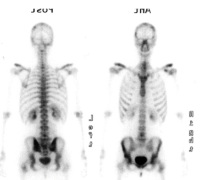
Radionuclide scan showing extensive skeletal involvement with diffuse
vertebral and iliac bilateral localizations. Keywords: radionuclide scan,
skeletal involvement
|
|
|
7. Pace A;
Villani V
Extra-Cranial Metastases from Glioblastoma Multiforme
Spine
European Association of NeuroOncology Magazine 2012; 2 (2): 91-92
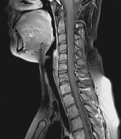
Cervical spine T1-weighted MRI image with
gadolinium showing a mild leptomeningeal enhancement. Keywords: leptomeningeal enhancement,
MRI,
spine
|
|
|
8. Pace A;
Villani V
Extra-Cranial Metastases from Glioblastoma Multiforme
Spine
European Association of NeuroOncology Magazine 2012; 2 (2): 91-92
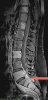
Spine T1-weighted MRI image with gadolinium.
The red arrow indicates the epidural tissue in the epidural space at the lumbosacral level. Keywords: lumbo-sacral level,
MRI,
spine
|
|
|
9. Pace A;
Villani V
Extra-Cranial Metastases from Glioblastoma Multiforme
Spine
European Association of NeuroOncology Magazine 2012; 2 (2): 91-92
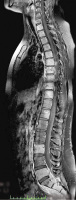
Spine T1-weighted MRI image fat-sat
showing a diffuse signal alteration in the vertebral
bodies plus the presence of tissue at the
lumbo-sacral level. Keywords: lumbo-sacral level,
MRI,
spine
|
|
|
10. Dittrich M;
Miederer SE;
Havertz B;
Krastev R
Schaumzerstörung und Schaumverhinderung: Der Wirkmechanismus von Simeticon in vitro
Antifoaming
Journal für Gastroenterologische und Hepatologische Erkrankungen 2010; 8 (3): 19-25
Antifoaming: Zeiten bis zum ersten Sichtbarwerden einer C12G2-Lösung
nach Zugabe von Simeticon verschiedener Konzentrationen aus LPL, Lefax Kautabletten
und Lefax Kautabletten extra. Man beachte die doppelt logarithmische
Skalierung. |
|
|
11. Dittrich M;
Miederer SE;
Havertz B;
Krastev R
Schaumzerstörung und Schaumverhinderung: Der Wirkmechanismus von Simeticon in vitro
Simeticon
Journal für Gastroenterologische und Hepatologische Erkrankungen 2010; 8 (3): 19-25
Oberflächenspannungen von Simeticon in LPL, Lefax Kautabletten und Lefax Kautabletten extra in verschiedenen Verdünnungen in Wasser. |
|
|
12. Tamse CT;
Lu X;
Mortel EG;
Cabrales E;
Feng W;
Schaefer S
The Peripheral Benzodiazepine Receptor Modulates Ca2+Transport through the VDAC in Rat Heart Mitochondria
Calcium Kinetics
Journal of Clinical and Basic Cardiology 2008; 11 (1-4): 24-29
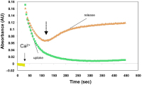
A representative tracing of extra-mitochondrial calcium kinetics following the addition of 60 μM Ca2+ into a solution containing isolated cardiac mitochondria and the calcium-sensitive marker
APIII. The absorbance (AU) increased rapidly with the addition of Ca2+ (solid arrow), followed by a decline representing uptake of calcium
by mitochondria. The slope and magnitude of this decline was quantified as calcium uptake rate and amount. Following mitochondrial loading with calcium, an increase in AU reflected calcium release
by mitochondria (dashed arrow). Calcium release was quantified as the slope of this line, and was partially inhibited by CsA. The lower trace (no hemin) shows rapid uptake of calcium and an absence of release following loading. The upper trace (hemin = 10 μM) illustrates a reduction in calcium loading as well as early release. Keywords: calcium kinetics,
flowchart
|
|
|
13. Bergmann P;
Huber S;
Keeling I;
Martin D;
Mächler H;
Oberwalder PJ;
Rigler B
Neue Anastomosendevices in der Herzchirurgie
Anastomosendevices
Journal für Kardiologie - Austrian Journal of Cardiology 2004; 11 (4): 153-157
Das Ventrica-Device zur Fertigung distaler Magnetanastomosen (A: Plazierungsgriff,
B: Auslöser, C: Positionierungsgriff, D: Gehäuse für Magnetclips, E: extra- und intravaskuläre Magnetclips) Keywords: Anastomose,
Device,
Foto,
Kardiologie,
Magnetanastomose,
Ventrica-Device
|
|
|
14. Stauber R
Management chronischer Hepatopathien - State of the art 2003
Primärsklerosierende Cholangitis
Journal für Gastroenterologische und Hepatologische Erkrankungen 2003; 1 (1): 21-27
ERCP bei einem 40jährigen Patienten mit primär-sklerosierender Cholangitis (PSC). Die Gallengänge wurden über einen okkludierenden Ballonkatheter mit Kontrastmittel aufgespritzt. Die extra- und intrahepatischen Gallengänge sind durch hochgradige Strikturen und dazwischenliegende zystische Erweiterungen völlig destruiert. Klinisch bestand Ikterus (Bilirubin um 20 mg/dl) und massiver cholestatischer Pruritus; mittlerweile ist der Patient erfolgreich lebertransplantiert. Keywords: Endoskopische retrograde Cholangiopankreatographie,
ERCP,
Gallengang,
Hepatologie,
Primärsklerosierende Cholangitis
|
|
|
15. Gasser R;
Gasser S;
Hecking C
Ultraweak Electromagnetic Fields and Sub-Atomic Dynamics - A Possible Subset of Signal-Transduction and -Storage Mechanisms in the Cardiovascular System (Non-Linearity, Small Scale Fluctuations and Predictive Aspects
Leitfähigkeit - Magnesium
Journal of Clinical and Basic Cardiology 2003; 6 (1-4): 87-92
Results from own voltage-clamp experiment using isolated cardiac sheep Purkinje strands, where the slope conductance increases when extra-cellular Mg (Mgo) is reduced. The isochronal current-voltage relationship under control conditions (filled triangles) changes after reducing Mgo to near zero (open triangles) and the outward current increases at depolarised potentials. The presence
of 0.3 mM barium decreases the slope conductance to the same values regardless of the presence (closed squares) and absence of Mgo (open squares). Inset: open circles: Bao-sensitivity of currentvoltage relationship in the absence of Mgo, ie, the difference between Mgo sensitive current and Barium-inhibited current-voltage relationship
at near 0 Mgo; filled circles: Mgo-sensitive current-voltage relationship, ie, difference between control and the current-voltage relationship at Mgo near 0 mM. It can be seen clearly that changing concentrations of extracellular divalent cation concentrations affect transmembrane conductance through voltage gated ion channels. Keywords: Barium,
Barium,
conductance,
diagram,
Diagramm,
Leitfähigkeit,
magnesium,
magnesium,
Purkinje strand,
Purkinje-Fasern,
Spannung,
voltage
|
|
|
16. Gasser R;
Horn S;
Köppel H
Discrimination by valinomycin K-selective surface microelectrodes of a sulphonylurea-sensitive and a distinct sulphonylurea-, barium-, TEA- and cinnamate-insensitive component of K-efflux from isolated pig coronary arteries during simulated ischaemia
K-Efflux in Koronararterien - Mikroelektroden
Journal of Clinical and Basic Cardiology 1998; 1 (1): 43-51
Simplified diagram of different microelectrode tips during simulated ischaemia. Hatched area represents paraffin oil (P.o.), area underneath, marked with oval rings, represents myocardial or smooth muscle tissue (T). Between tissue and P.o. one can see the artificially created reduced extra-cellular space (ARECS). a. Electrode tip bevelled in a 45° angle not yielding ideal contact with the tissue surface, thus making contact with the paraffin oil. Under these conditions the hydrophobic carrier will instantly escape from the tip and the electrode does not give further readings. b. Elec-trode tip bevelled in a 45° angle pushed too deeply into the tissue causing cell damage. Cells will be
either damaged or destroyed causing a constant leak of K+(K+ ions represented by stippling) into the ARECS which, in turn, leads to faulty measurements: instead of ischaemia-induced K+ efflux caused by changed K+ conductance of the membrane the electrode measures changes resulting from damaged cells (c.f. Fig. 3). c. Electrode tip bevelled in a 90° angle (flat tip) ideally positioned in the ARECS. Only under these circumstances correct measurements of transmembrane K+ efflux induced by ischaemia are possible (c.f. Fig. 3). d. Ideally bevelled surface microelectrode, impaled too deeply into the tissue. Stippling represents K+ ions leaking from damaged tissue - readings will be misleading. e. Too fine-tipped microelectrode, pushed too deeply into the tissue. Leaking K+ from the damaged tissue like in b and d, represented by stippling and small arrows. Keywords: Arteria coronaria,
Arteria coronaria,
Efflux,
Efflux,
Graphische Darstellung,
ischaemia,
Ischämie,
Kalium,
microelectrode,
Mikroelektrode,
potassium
|
|
|
| |

 Suche Medizinische Publikationen
 Suche Abbildungen und Grafiken
 Medizinprodukte - Labortechnik
 Antidotarium (Stand 2012)
 Phytokodex (Stand 2012)
 Fachbücher
 Patientenliteratur