|
|
|---|
Oberbegriffe: Sion
Unterbegriffe: AMI-Perfusion,
Anisotrope Diffusion,
Beinperfusion,
Bildfusion,
Bildfusionierung,
CMR-Perfusionsuntersuchung,
CT-Fusion,
Diffusion,
Diffusion-Perfusion-Impairment,
Diffusiongewichtung,
Diffusions-MRT,
Diffusionsgewichtete,
Diffusionskoeffizient,
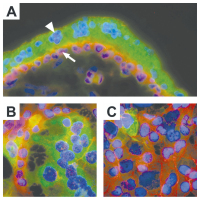
Diffusionsstörung,
Diffusionstensors,
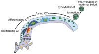
First-Pass-Perfusion,
First-Pass-Perfusionsstudie,
Fusionierte,
Fusionierung,
Fusions-FDG-PET-CT
Verwandte Begriffe: AV-Knotendepression,
Ablationsläsionen,
Accession,
Adhesion,
Adhäsion,
Adhäsions-Rezeptoren,
Adhäsionsbildung,
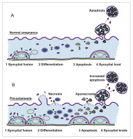
Aggression,
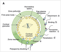
Aggressionsverhalten,
Ammoniak-Positronenemissionstomographie,
Ammoniak-Positronenemmissionstomographie,
Androgensuppression,
Aortenläsion,
Aquaporinexpression,
Arrosion,
Arrosionsblutung,
Arzneimittelkommission,
BG-Läsion,
Ballonokklusion,
Ballonokklusionssystems
|
1. Kröll M;
Strouhal A;
Fiegl N
Aortenklappeninsuffizienz: Was gibt es Neues? // Aortic valve regurgitation – What do the 2021 guidelines say? What has changed from 2017?
Echo
Journal für Kardiologie - Austrian Journal of Cardiology 2024; 31 (5-6): 120-128
3D-TEE, bikuspide Aortenklappe in der Systole, Fusion von
rechts- und linkskoronarer Tasche mit sklerosierter Raphe. Keywords: Echo,
Kardiologie
|
|
|
2. Neuhold U
Empfehlungen zur CRT-Nachsorge // Recommendations for CRT
EKG
Journal für Kardiologie - Austrian Journal of Cardiology 2023; 30 (9-10): 220-231
(a): QRS-Dauer 116 ms; Pacing-Modus: Fusion LV-Pacing mit intrinsischer Leitung über
den rechten Tawaraschenkel. (b): QRS-Dauer 125 ms; Pacing-Modus: biV-Pacing. Keywords: EKG,
Kardiologie
|
|
|
3. Zweiker D;
Bisping E;
Lercher P;
Manninger M;
Prenner G;
Riedlbauer R;
Scherr D
EPU-Corner: Incessant WPW-Syndrom
EKG bei Aufnahme
Journal für Kardiologie - Austrian Journal of Cardiology 2018; 25 (5-6): 148-149
Elektrokardiogramm bei der Aufnahme auf die Intensivstation. Rhythmische Schmalkomplextachykardie, Frequenz 198/min, Rechtstyp, Fusion der P- und T-Wellen, QRS-Dauer 70 ms, R/S-Umschlag in V5, negative Ts in II, III, aVF, QT 270ms, QTc 402ms. Keywords: Aufnahme,
EKG,
Elektrokardiogramm,
Kardiologie,
WPW-Syndrom
|
|
|
4. Bellmann B;
Deppe I;
Nagel P;
Roser M;
Muntean BG
Case Report: Pacemaker Mimics Ventricular Extrasystoles?
Ventricular premature beats
Journal für Kardiologie - Austrian Journal of Cardiology 2017; 24 (11-12): 286-287
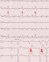
Resting ECG with recordings of ventricular premature beats with left bundle branch block morphology. These are marked by the red arrow and show a partial pseudo- fusion. Keywords: left bundle branch block,
Linksschenkelblock,
resting ECG,
Ruhe-EKG,
ventricular extrasystole,
ventricular premature beat,
ventrikuläre Extrasystole
|
|
|
5. Steichen-Gersdorf E;
Kapelari K;
Hofer S
Wachstumsstörung bei SHOX-Defizienz // SHOX Deficiency Disorders
Radiologische Zeichen
Journal für Klinische Endokrinologie und Stoffwechsel - Austrian Journal of Clinical Endocrinology and Metabolism 2016; 9 (3): 67-70
Radiologische Zeichen der SHOX-Defizienz, Knochenalter 13 Jahre a) Fusion der ulnaren Radiusepiphyse (Triangulation), siehe Pfeil b) Aufhellung der distalen Radiusmetaphyse (Radiolucency), *
c) spitzer Karpalwinkel (Pyramidalisation der proximalen karpalen Reihe) Keywords: radiologische Zeichen,
Radiusepiphyse,
Radiusmetaphyse,
Röntgen,
SHOX-Defizienz,
Triangulation
|
|
|
6. Götz I;
Grosu AL;
Spehl TS
Role of PET Imaging in Patients with High-Grade Gliomas undergoing anti-angiogenic Therapy with Bevacizumab – Review of the Literature and Case Report
Glioblastoma
European Association of NeuroOncology Magazine 2014; 4 (3): 102-108
Imaging results of a patient with glioblastoma undergoing bevacizumab
therapy. First column: MRI (MP-RAGE with contrast enhancement); second column:
18F-FET-PET (parametric images scaled to mean uptake in a right temporal reference
region); third column: image fusion. (A) scan prior to therapy 07/2010; (B) scan after
8 weeks of bevacizumab 09/2010; (C) scan after 16 weeks of bevacizumab 11/2010;
(D) scan after re-irradiation with 20 Gy, 01/2011. Bevacizumab therapy was continued.
For details, see text Keywords: Bevacizumab,
glioblastoma,
MRI
|
|
|
7. Schwabl C;
Gaugg M;
Beham-Schmid C;
Celedin S;
Liegl-Atzwanger B;
Matschnig S;
Raunik W;
Weiss H;
Neumann HJ
Rheumatoide Arthritis und seltene Tumormanifestation – auf Umwegen zur Remission
Sarkom
Tumorboard 2014; 3 (1): 5-9
Fusions-FDG-PET-CT (06/2010): Rechts paravertebral beginnend an der
Aortenbifurkation bis prävertebral von S5 mäßig FDG-positive Läsion – suspekt auf
einen Weichteiltumor. Keywords: CT,
FDG-PET,
Onkologie,
Rezidiv,
Sarkom,
Weichteiltumor
|
|
|
8. Perathoner A;
Kafka-Ritsch R;
Eisterer W;
Bechter O;
Pall G;
Zitt M
Interdisziplinär-multimodale Behandlung einer jungen Frau mit einem Desmoplastic Small Round Cell Tumor (DSRCT), einem sehr seltenen und aggressiven Tumor des Peritoneums
DSRCT
Tumorboard 2013; 2 (1): 17-22
PET-CT- Fusion mit positivem Tumorknoten im Unterbauch (1. Rezidiv) Keywords: DSRCT,
Onkologie,
PET-CT,
Rezidiv,
Tumorknoten
|
|
|
9. Proescholdt M;
Doenitz C;
Brawanski A
Surgery of Malignant Gliomas Using Modern Technology
Glioma - Surgery
European Association of NeuroOncology Magazine 2013; 3 (1): 11-14
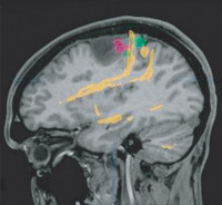
Fusion of [F-18]fluoroethyltyrosine: PET (red), functional MRI (green), and
DTI tractography (yellow) in a patient with a left frontal anaplastic astrocytoma.
Note the close vicinity of eloquent cortical and subcortical structures to the tumour
borders. Keywords: glioma,
MRI,
PET
|
|
|
10. Decristoforo I;
Maier H;
Gugl K
Stabilisierende Operationen an der Wirbelsäule: Ein Überblick
LWS
Journal für Neurologie, Neurochirurgie und Psychiatrie 2011; 12 (1): 32-37
(a) Nativröntgen präoperativ anterior/posterior und seitlich; (b) Nativröntgen präoperativ seitlich; (c–e) CT der LWS präoperativ; Kontrolle ein Jahr nach Dekompression
L2–L4 und ventrodorsaler Fusion L1–L5: uneingeschränkte Gehstrecke, keine LWS-Schmerzen, bleibt in Kontrolle; (f) Nativröntgen anterior/posterior; (g) Nativröntgen
seitlich: Wiederherstellung der Lordose. Keywords: Lordose,
LWS,
Neurochirurgie
|
|
|
11. Grimmer A;
Wurm G
Chirurgische Strategien bei Bandscheibenvorfällen an der Halswirbelsäule
HWS - MRT
Journal für Neurologie, Neurochirurgie und Psychiatrie 2011; 12 (1): 23-30
(a) Präoperatives HWS-MRT, sagittal in T2-Gewichtung: Bandscheibenvorfall
und Retrospondylose C5/C6 und C6/C7 mit konsekutiver Myelopathie und
absoluter Wirbelkanalstenose. (b) Postoperatives HWS-MRT, sagittal in T2-Gewichtung:
nach „Anterior Cervical Discectomy and Fusion with Instrumentation“ (ACDFI)
mit ventraler winkelstabiler Platte und 2 PEEK- (Polyetheretherketon-) Cages auf
Höhe C5/C6 und C6/C7. (c) Postoperatives HWS-Röntgen anterior/posterior: Mit
ventraler Platte und 2 PEEK-Cages. (d) Postoperatives seitliches HWS-Röntgen. Keywords: HWS,
MRT,
Neurochirurgie
|
|
|
12. Grimmer A;
Wurm G
Chirurgische Strategien bei Bandscheibenvorfällen an der Halswirbelsäule
HWS
Journal für Neurologie, Neurochirurgie und Psychiatrie 2011; 12 (1): 23-30
Operative Zugangswege zur Halswirbelsäule. ACDF: Anterior Cervical Discectomy and Fusion; ACD: Anterior Cervical Discectomy; ACDFI: Anterior Cervical Discectomy and Fusion with Instrumentation. Keywords: HWS,
Neurochirurgie,
Operation
|
|
|
13. Felber SR
Radiologische Diagnostik bei Wirbelsäulenerkrankungen
CT - Wirbelsäule
Journal für Neurologie, Neurochirurgie und Psychiatrie 2011; 12 (1): 9-14
Zustand nach ventraler Fusion HWK 4/5 und
HWK 5/6 und Zustand nach Implantation einer Bandscheibenprothese
HWK 3/4 in der seitlichen Röntgenaufnahme
(a). Die Bandscheibenprothese verursacht am 3-Tesla-MRTGerät
(b) ein Artefakt, das die Beurteilung des Rückenmarks
einschränkt. Am offenen 1-Tesla-MRT (c–e) stört das Metallartefakt
die Beurteilung nicht. In den funktionellen Aufnahmen
in Reklination (d) und Inklination (e) besteht ausreichende
Beweglichkeit der Halswirbelsäule und es zeigt sich
keine Einengung des Spinalkanals. Das Rückenmark weist
eine zentrale Läsion im Sinne der zervikalen Myelopathie auf. Keywords: CT,
Wirbelsäule
|
|
|
14. Martinek M;
Meyer Ch;
Aichinger J;
Nesser H-J;
Pürerfellner H
Die Entwicklung der Radiofrequenzablation von Vorhofflimmern in den Jahren 2000 bis 2009 am Beispiel der Elektrophysiologie der Elisabethinen Linz
Visualisierung
Journal für Kardiologie - Austrian Journal of Cardiology 2010; 17 (11-12): 412-418
Entwicklung der Visualisierung: (a) Segmental-ostiale Pulmonalvenenisolation
(fluoroskopisch geführt); durchgezogener Pfeil: Ablationskatheter;
unterbrochener Pfeil: zirkulärer diagnostischer Katheter; gepunkteter Pfeil: Koronarsinuskatheter.
Zirkumferenzielle Pulmonalvenenisolation: (b) Elektroanatomisches
Map (CARTO); (c) Fusion von Elektroanatomischem Map und Multislice-Computertomographie
(CARTOMerge); (d) Fast Anatomical Map (CARTO 3) Keywords: Kardiologie,
Radiofrequenzablation,
Visualisierung
|
|
|
15. Kürzl R
Differentialdiagnose von Hautveränderungen der Vulva
Vulva - Lichen
Speculum - Zeitschrift für Gynäkologie und Geburtshilfe 2008; 26 (2) (Ausgabe für Österreich): 12-15
Speculum - Zeitschrift für Gynäkologie und Geburtshilfe 2008; 26 (2) (Ausgabe für Schweiz): 12-12
Ausgedehnte erythroplakische Hautveränderung mit fast vollständiger Rückbildung der kleinen Labien
(links vorne noch rudimentär erhalten)
und mit Fusion des Präputium klitoridis bei einer 71-jährigen Patientin. Histologie: Lichen ruber
planus. Keywords: Foto,
Gynäkologie,
Lichen ruber planus,
Speculum,
Vulva
|
|
|
16. Kürzl R
Differentialdiagnose von Hautveränderungen der Vulva
Vulva - VIN
Speculum - Zeitschrift für Gynäkologie und Geburtshilfe 2008; 26 (2) (Ausgabe für Österreich): 12-15
Speculum - Zeitschrift für Gynäkologie und Geburtshilfe 2008; 26 (2) (Ausgabe für Schweiz): 12-12
Ausgedehnte erythroplakische Hautveränderung mit vollständiger Rückbildung der kleinen Labien
und mit Fusion des Präputium klitoridis bei einer 76-jährigen Patientin. Histologie: Vulväre
intraepitheliale Neoplasie (VIN) Keywords: Foto,
Gynäkologie,
Speculum,
Vulva,
Vulväre intraepitheliale Neoplasie
|
|
|
17. Gauster M;
Huppertz B
Fusion of Cytothrophoblast with Syncytiotrophoblast in the Human Placenta: Factors Involved in Syncytialization
Trophoblast fusion
Journal für Reproduktionsmedizin und Endokrinologie - Journal of Reproductive Medicine and Endocrinology 2008; 5 (2): 76-82
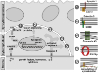
Illustration of factors involved in trophoblast fusion. Environmentally derived growthfactors, hormones and cytokines bind to their cognate receptors at the plasma membrane ofcytotrophoblasts. Activation of either protein kinase A (PKA) or MAP kinases ERK1/2 and p38leads to increased protein expression of the transcription factor GCMa, which in turn drivestranscription of fusogenic genes. Several structural and membrane proteins were suggested topromote trophoblast fusion, including syncytin 1 and its receptor ASCT2 (1), CD98 and its receptorgalectin 3 (2) as well as connexin 43 (3). Beside activation of PKA or MAP kinase pathways,cytokines induce conversion of pro-caspase 8 into active caspase 8. Once activated, caspase 8can mediate inactivation of “flippasesâ€� and/or activation of "floppases" to trigger phosphatidyl-serine externalisation (PS flip) (4). Additionally, caspase 8 triggers remodelling of the sub-mem-branous cytoskeleton by degrading structural proteins such as α-fodrin (5). Keywords: Throphoblast,
trophoblast fusion
|
|
|
18. Gauster M;
Huppertz B
Fusion of Cytothrophoblast with Syncytiotrophoblast in the Human Placenta: Factors Involved in Syncytialization
Human placenta
Journal für Reproduktionsmedizin und Endokrinologie - Journal of Reproductive Medicine and Endocrinology 2008; 5 (2): 76-82
A) Immunofluorescent staining ofhuman first trimester placenta (week 8).Mononucleated cytotrophoblasts (arrow)show staining for E-cadherin in their apicaland lateral cell membranes (red). The over-lying multinucleated syncytiotrophoblast(arrowhead) lacks lateral cell walls and isnegative for E-cadherin. The syncytiotro-phoblast expresses hCG (green). B) and C)Immunofluorescent staining of forskolin andvehicle treated BeWo cells. Treatment ofBeWo cells for 48h with forskolin (20 μM)induced formation of multinucleated syn-cytia, visualized by degradation of E-cadherin (stained in red). hCG expression(green) was predominantly observed in syn-cytia of forskolin treated BeWo cells, butwas also rarely seen in mononucleatedBeWo cells (treated and untreated). Keywords: placenta,
Plazenta
|
|
|
19. Gauster M;
Huppertz B
Fusion of Cytothrophoblast with Syncytiotrophoblast in the Human Placenta: Factors Involved in Syncytialization
Trophoblast
Journal für Reproduktionsmedizin und Endokrinologie - Journal of Reproductive Medicine and Endocrinology 2008; 5 (2): 76-82
Schematic representation of trophoblast differentiation within the villous trophoblast.Derived from trophoblast progenitor cells daughter cells start to differentiate and finally fusewith the overlying syncytiotrophoblast. Nuclei that have become an integrative part of the syn-cytiotrophoblast change their morphology and display chromatin condensation. Finally lateapoptotic syncytial nuclei are packed into syncytial knots and are released into the maternalblood stream. The arrows follow the route of cell types and nuclear changes during trophoblastdifferentiation. Keywords: Schema,
scheme,
Trohoblast,
trophoblast
|
|
|
20. Röhl K;
Weidt W;
Röhrich F
Moderne Behandlung der zervikalen Diskopathie bei Querschnittlähmung
HWS
Journal für Mineralstoffwechsel & Muskuloskelettale Erkrankungen 2008; 15 (2): 71-73
Die Funktionsaufnahmen der HWS zeigen bei Z. n. Fusion der Etagen HWK 4/5/6 eine Anschlussinstabilität im Segment HWK 3/4. Keywords: HWS,
Mineralstoffwechsel,
Röntgenbild
|
|
|
21. Becker S;
Meissner J;
Bretschneider W;
Bartl R;
Ogon M
Stabilisierende Operationen an der Wirbelsäule bei schwerer Osteoporose
Osteoporose
Journal für Mineralstoffwechsel & Muskuloskelettale Erkrankungen 2007; 14 (3): 107-112
80jährige Patientin mit Fraktur Th11: (a): Zunehmende neurologische Ausfälle und Claudicatio spinalis. (b): Versorgung mit Dekompression, offener Ballonkyphoplastie und dorsaler Fusion Th9 bis L1 mit zusätzlicher dorsaler Knochenanlagerung (Pfeil). Keywords: Fallbericht,
Mineralstoffwechsel,
Röntgenbild,
Wirbelsäule
|
|
|
22. Somay C
Fallbericht: Bicalutamid 150 mg (Casodex) als Salvage-Therapie beim Lokalrezidiv nach Permanentimplantation der Prostata mit J-125-Seeds
F18-Cholin-PET mit CT-Fusion
Journal für Urologie und Urogynäkologie 2007; 14 (2) (Ausgabe für Österreich): 20-22
F18-Cholin-PET mit CT- Fusion (Lokalrezidiv mit Pfeil gekennzeichnet) Keywords: PET,
PET,
Positron emission tomography,
Positronenemissionstomographie
|
|
|
23. Steinwender C;
Hönig S;
Hofmann R;
Patrasso M;
Kneidinger R;
Leisch F
Fusionierung von Geometrie und CT (Pulmonalvenenisolation ohne Spiralkatheter)
CT - Virtuelle Anatomie
Journal für Kardiologie - Austrian Journal of Cardiology 2007; 14 (Supplementum C): 7-9
Beginn der Fusion zwischen CT- und virtueller Anatomie (posterioranterior). Keywords: Kardiologie,
linker Vorhof
|
|
|
24. Martinek M;
Aichinger J;
Nesser H-J;
Böhm G;
Pürerfellner H
Fusion von CT und 3D-Elektroanatomie zur individualisierten Ablation von Vorhofflimmern - das CartoMerge TM Image Integration Module
Vorhofflimmern - Fallbericht
Journal für Kardiologie - Austrian Journal of Cardiology 2006; 13 (9-10): 306-310
Bearbeitete CT-Darstellung; posterior-anteriore Ansicht (Blau = Ösophagus;
Grün = linkes Atrium; Rot = Sinus coronarius; Gelb = linkes Herzohr). Keywords: 3D-Mapping,
Fallbericht,
Kardiologie,
Vorhofflimmern
|
|
|
25. Martinek M;
Aichinger J;
Nesser H-J;
Böhm G;
Pürerfellner H
Fusion von CT und 3D-Elektroanatomie zur individualisierten Ablation von Vorhofflimmern - das CartoMerge TM Image Integration Module
Vorhofflimmern - Fallbericht
Journal für Kardiologie - Austrian Journal of Cardiology 2006; 13 (9-10): 306-310
Bearbeitete CT-Darstellung; linke schräge Ansicht (Grün = Sinus coronarius, Orange = linkes Atrium, Rot = linkes Herzohr, Türkis = Ösophagus) Keywords: 3D-Mapping,
Fallbericht,
Kardiologie,
Vorhofflimmern
|
|
|
26. Martinek M;
Aichinger J;
Nesser H-J;
Böhm G;
Pürerfellner H
Fusion von CT und 3D-Elektroanatomie zur individualisierten Ablation von Vorhofflimmern - das CartoMerge TM Image Integration Module
Vorhofflimmern - Fallbericht
Journal für Kardiologie - Austrian Journal of Cardiology 2006; 13 (9-10): 306-310
Rot = Aortenwurzel mit den Abgängen der Koronargefäße (schwarzer
Pfeil = Ramus circumflexus der linken Kranzarterie); Gelb = linkes Atrium; Orange =
linkes Herzohr; Grün = linker Ventrikel; Dunkelblau = Pulmonalarterien-Hauptstamm;
Hellblau = Ösophagus; linke schräge Ansicht (Patientenbeschreibung siehe Abb. 2). Keywords: 3D-Mapping,
Fallbericht,
Kardiologie,
Vorhofflimmern
|
|
|
27. Martinek M;
Aichinger J;
Nesser H-J;
Böhm G;
Pürerfellner H
Fusion von CT und 3D-Elektroanatomie zur individualisierten Ablation von Vorhofflimmern - das CartoMerge TM Image Integration Module
Vorhofflimmern - Fallbericht
Journal für Kardiologie - Austrian Journal of Cardiology 2006; 13 (9-10): 306-310
62jährige Patientin mit persistierender Vorhofflimmerarrhythmie seit 6 Monaten mit konsekutiver Belastungsdyspnoe und Zustand nach mehrmaliger elektrischer Kardioversion; Klasse-1c-Antiarrhythmikum ineffektiv, Sedacoron-Unverträglichkeit bei zusätzlicher immunogener Hyperthyreose und Zustand nach Radiojodtherapie;
zirkumferentielle Ablation inklusive mitraler Isthmuslinie; seit der Ablation
keine Rezidive (subjektiv und in mehreren Langzeit-EKGs) ohne antiarrhythmische
Medikation über 5 Monate. CartoMerge™-Map nach zirkumferentieller Ablation, posterior-anteriore Ansicht (Rot = Ablationspunkte; Hellgelb = CT-Bild; Türkis = darunter liegendes CartoMerge™-Map; durchscheinend Blau = Ösophagus; "dach" =
definierte Bezugspunkte für die Bild fusionierung) Keywords: 3D-Mapping,
Computertomographie,
Fallbericht,
Kardiologie,
Vorhofflimmern
|
|
|
28. Martinek M;
Aichinger J;
Nesser H-J;
Böhm G;
Pürerfellner H
Fusion von CT und 3D-Elektroanatomie zur individualisierten Ablation von Vorhofflimmern - das CartoMerge TM Image Integration Module
CT-Bild - CartoMergeTM-Map
Journal für Kardiologie - Austrian Journal of Cardiology 2006; 13 (9-10): 306-310
Fusionierung des CT-Bildes (Grau) und des elektroanatomischen CartoMerge™-Maps (Türkis bzw. als Gitternetz mit einzelnen Punkten); "Roof" = definierte Bezugspunkte für die Bildfusionierung; a) anterior-posteriore Ansicht, b) posterior-anteriore Ansicht. Keywords: 3D-Mapping,
Computertomographie,
Kardiologie
|
|
|
29. Martinek M;
Aichinger J;
Nesser H-J;
Böhm G;
Pürerfellner H
Fusion von CT und 3D-Elektroanatomie zur individualisierten Ablation von Vorhofflimmern - das CartoMerge TM Image Integration Module
Vorhofflimmern - Fallbericht
Journal für Kardiologie - Austrian Journal of Cardiology 2006; 13 (9-10): 306-310
60jähriger Patient mit paroxysmalem Vorhofflimmern seit 15 Monaten und Zustand nach dreimaliger elektrischer Kardioversion; Ineffektivität einer Kombination von Betablocker und Klasse-Ic-Antiarrhythmikum; nach zirkumferentieller Pulmonalvenenisolation inklusive Dachlinie seit nunmehr 6 Monaten unter Betablockertherapie rezidivfrei. Bearbeitetes CT-Bild und elektroanatomisches Carto™-Map vor der Fusionierung; a) anterior-posteriore Ansicht; b) LAO-Ansicht; c) posterior-anteriore Ansicht. Keywords: 3D-Mapping,
Computertomographie,
Fallbericht,
Kardiologie,
Vorhofflimmern
|
|
|
30. Martinek M;
Aichinger J;
Nesser H-J;
Böhm G;
Pürerfellner H
Fusion von CT und 3D-Elektroanatomie zur individualisierten Ablation von Vorhofflimmern - das CartoMerge TM Image Integration Module
Vorhofflimmern - Fallbericht
Journal für Kardiologie - Austrian Journal of Cardiology 2006; 13 (9-10): 306-310
69jähriger Patient mit paroxysmalem Vorhofflimmern seit 15 Jahren und zuletzt persistierendem Vorhofflimmern seit 4 Monaten; Zustand nach mehrmaliger elektrischer Kardioversion; Ineffektivität mehrerer Klasse-Ic- und Klasse-III-Antiarrhythmika; nach zirkumferentieller Pulmonalvenenisolation inklusive Dachlinie seit nunmehr 7 Monaten ohne Antiarrhythmikum rezidivfrei. Bearbeitete CT-Bilder (Gelb = linkes Atrium, Rot = linkes Herzohr, Dunkelblau, durchscheinend = Ösophagus; Blauviolett = Pulmonalvenen);
a) anterior-posteriore Ansicht; b) LAO-Ansicht; c) posterior-anteriore Ansicht. Keywords: 3D-Mapping,
Computertomographie,
Fallbericht,
Kardiologie,
Vorhofflimmern
|
|
|
31. Huppertz B
Placental Villous Trophoblast: the Altered Balance Between Proliferation and Apoptosis Triggers Pre-eclampsia
Trophoblast turnover
Journal für Reproduktionsmedizin und Endokrinologie - Journal of Reproductive Medicine and Endocrinology 2006; 3 (2): 103-108
Schematic representation of trophoblast turnover. (A) Normal pregnancy: The final event of cytotrophoblast differentiation, syncytial fusion, results in incorporation of fresh organelles and other cellular
material into the syncytium (1). Within the multinucleated syncytiotrophoblast
differentiation (2) and subsequently late apoptosis (3) take place. Finally, the late apoptotic material is packed into protrusions of the apical plasma membrane, syncytial knots. These knots are released into the maternal circulation as tightly sealed corpuscles (4). (B) Pre-eclampsia: Enhanced proliferation and syncytial fusion (1) may overwhelm the
capacity of the syncytiotrophoblast in terms of differentiation (2) and
apoptotic release (3). This may result in a necrotic breakdown of specific
sites of the syncytiotrophoblast. If at these sites apoptosis has not yet started, pure necrosis can be observed; if however apoptosis has already lead to first cleavage of proteins, apoptotic material will be necrotically released (aponecrosis). At the same time the syncytiotrophoblast tries to counter balance for the increased input by increasing the release of apoptotic syncytial knots (4). Modified from [15]. Keywords: pregnancy,
Schema,
scheme,
Schwangerschaft,
trophoblast
|
|
|
32. Koyama K;
Hasegawa A
Premature Ovarian Failure Syndrome May Be Induced by Autoimmune Reactions to Zona Pellucida Proteins
Zona pellucida
Journal für Reproduktionsmedizin und Endokrinologie - Journal of Reproductive Medicine and Endocrinology 2006; 3 (2): 94-97
Role of the zona pellucida (ZP) in fertilisation and oocyte maturation. A: Schematic drawing of sperm-ZP interaction (modified from [11]). The capacitated sperm attaches and then tightly binds to the ZP (a, b). These interactions are mediated by ZP3 which serves as a primary receptor
for sperm. The acrosome reaction in the bound sperm is induced which is essential for sperm penetration of ZP (c). ZP2 maintains acrosomereacted sperm in the ZP during penetration (d). Following fusion of egg and sperm, cortical granules disintegrate to induce the zona reaction that prevents polyspermy (e, f). Proteolytic cleavage of ZP2 and ZP3 is involved in the zona reaction. After fertilisation ZP still remains to protect the fertilised eggs. Keywords: Fertilisation,
oocyte maturation,
Oozyte,
Schema,
scheme,
Zona pellucida
|
|
|
33. Ogon M;
Becker S;
Chavanne A;
Meissner J;
Tuschel A
Die Bandscheibenprothese an der Halswirbelsäule
Bandscheibenprothese - Schmerz
Journal für Mineralstoffwechsel & Muskuloskelettale Erkrankungen 2006; 13 (1): 14-17
Klinische Frühergebnisse zeigen eine etwas raschere Beschwerdebesserung bei der Bandscheibenprothese (Bryan) im Vergleich zur monosegmentalen Fusion bei gleichem Ergebnis nach 12 Monaten. Keywords: Bandscheibenprothese,
Diagramm,
Mineralstoffwechsel,
Schmerz
|
|
|
34. Pürerfellner H;
Aichinger J;
Martinek M;
Nesser H-J;
Janssen J
Vergleich zweier Ablationsstrategien in der Behandlung von therapierefraktärem Vorhofflimmern - Entwicklung und Ergebnisse über einen 5-Jahreszeitraum
Elektroanatomie - CT-Anatomie
Journal für Kardiologie - Austrian Journal of Cardiology 2006; 13 (11-12): 336-343
Fusion von Elektroanatomie und dreidimensionaler CT-Anatomie (CartoMerge); posterior-anteriore Ansicht des linken Atriums und der Pulmonalvenen
nach Ablation (beige: CT-Darstellung linkes Atrium inkl. Pulmonalvenen; türkis:
elektroanatomisches Map; violett: Ösophagus; rot: Ablationspunkte) Keywords: 3D-Mapping,
Computertomographie,
CT,
Kardiologie,
linkes Atrium,
Pulmonalvene
|
|
|
35. Zech N
Plasticity of Stem Cells: Cell-fusion Versus Transdifferentiation
Adult stem cell plasticity
Journal für Reproduktionsmedizin und Endokrinologie - Journal of Reproductive Medicine and Endocrinology 2005; 2 (4): 239-245
Commonly used methods to examine adult stem cell plasticity and to discriminate fusion
from plasticity: cells of transgenic mice expressing markers such as green fluorescent protein (GFP)
or LacZ, which are expressed in all cells of transgenic mice, are frequently used to demonstrate the plasticity potential of certain cells, originally belonging to a given cell lineage. For example, when GFP-positive bone marrow cells are injected into wildtype mice and these cells are found in tissues not related to the tissue of origin, it is believed that a lineage switch has occurred. In the case of transplantation of bone marrow cells, lethal irradiation of the recipient should guarantee that no stem cells of the wild type mouse contributes to the observed transdifferentiation or fusion. Keywords: plasticity,
Plastizität,
Reproduktionsmedizin,
Schema,
scheme,
Stammzelle,
stem cell
|
|
|
36. Zech N
Plasticity of Stem Cells: Cell-fusion Versus Transdifferentiation
Sex mismateched stem cell transplantation
Journal für Reproduktionsmedizin und Endokrinologie - Journal of Reproductive Medicine and Endocrinology 2005; 2 (4): 239-245
Sex mismatched stem cell transplantation to track progeny cells: claims on the plasticity of
stem cells often rely on the appearance of Y-positive cells in female recipients of male bone marrow transplant donors (left). Injection of marked bone marrow cells from female donors into male recipients is more suitable to demonstrate fusion events because marked Y chromosome-positive cells found in the recipient must have been formed by a fusion event (right). Keywords: Geschlecht,
Reproduktionsmedizin,
Schema,
scheme,
sex,
Stammzelle,
stem cell,
transplantation,
transplantation
|
|
|
37. Binder T
Three-Dimensional Echocardiography - Principles and Promises
3D-Echokardiographie
Journal of Clinical and Basic Cardiology 2002; 5 (2): 149-152
Three-dimensional reconstruction of a patient with mitral stenosis, morphologic features such as doming of the anterior leaflet and commissural fusion can be noticed. In addition, quantification of mitral valve area can be performed. Keywords: 3D,
3D,
echocardiography,
Echokardiographie,
Mitral valve,
Mitralklappe,
Stenose,
Stenosis
|
|
|
| |

 Suche Medizinische Publikationen
 Suche Abbildungen und Grafiken
 Medizinprodukte - Labortechnik
 Antidotarium (Stand 2012)
 Phytokodex (Stand 2012)
 Fachbücher
 Patientenliteratur