Content-type: text/html Suchergebnis - Abbildungen und Graphiken
Krause und Pachernegg
Verlag für Medizin und Wirtschaft
Mobile Version
A-  |   A  |   A+
Werbung
 
Suchergebnis - Abbildungen und Graphiken
 
Alle Journale wurden nach glioma surgery durchsucht Treffer 1 - 9 von 9

Medizinische Publikationen (10)  ::   Abbildungen und Graphiken  ::   Volltext (0)

Neue Suche:         Erweiterte Suche    Hilfe zur Suche

in allen Journalen nach  Bildtitel    Bildkeywords   Bildsummary   Autoren      Mindestens ein Wort  Alle Wörter   
1. Grossman R; Ram Z
Awake Craniotomy in Glioma Surgery
Awake Craniotomy
European Association of NeuroOncology Magazine 2014; 4 (1): 27-33
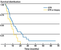
Kaplan-Meier curve of survival for elderly patients (65 years) with highgrade gliomas (HGG) who underwent awake-craniotomy gross tumour resection (GTR) compared to subtotal resection (STR) or biopsy. Median survival was significantly longer for patients with HGG who underwent GTR compared to those who underwent STR or biopsy (10.8 vs 7.8 months, respectively; p = 0.039) [39].

Keywords: awake craniotomy,  diagram,  glioma surgery,  Kaplan-Meier,  survival distribution
2. Grossman R; Ram Z
Awake Craniotomy in Glioma Surgery
Awake Craniotomy
European Association of NeuroOncology Magazine 2014; 4 (1): 27-33
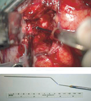
Sub-cortical stimulation of the cortico-spinal tracts by means of a subcortical monopolar electrode.

Keywords: awake craniotomy,  cortical stimulation,  glioma surgery,  photograph
3. Grossman R; Ram Z
Awake Craniotomy in Glioma Surgery
Awake Craniotomy
European Association of NeuroOncology Magazine 2014; 4 (1): 27-33
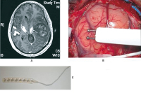
(A) Preoperative T1-weighted axial after gadolinium injection with diffuse tensor imaging (DTI) of the cortico-spinal tract showing a left temporo-parietal high-grade glioma displace medially the corticospinal tract. (B) Direct cortical stimulation by using bipolar probe. (C) A cortical strip electrode is placed over the surface of the motor cortex in order to assess motor-evoked potential (MEP).

Keywords: awake craniotomy,  cortical stimulation,  glioma surgery,  MRI,  photograph
4. Grossman R; Ram Z
Awake Craniotomy in Glioma Surgery
Awake Craniotomy
European Association of NeuroOncology Magazine 2014; 4 (1): 27-33
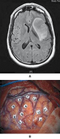
(A) Pre-operative axial fluid-attenuated inversion recovery- (FLAIR-) weighted MRI showing a WHO grade-II insular glioma in a 40-year-old woman presenting with episodes of seizures. This patient has no neurological deficit. (B) Intra-operative cortical mapping of language during awake surgery.

Keywords: awake craniotomy,  cortical mapping,  MRI,  photograph
5. Grossman R; Ram Z
Awake Craniotomy in Glioma Surgery
Awake Craniotomy
European Association of NeuroOncology Magazine 2014; 4 (1): 27-33
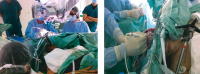
Intra-operative photographs of the surgical team during awake craniotomy.

Keywords: awake craniotomy,  glioma surgery,  photograph
6. Proescholdt M; Doenitz C; Brawanski A
Surgery of Malignant Gliomas Using Modern Technology
Glioma - Surgery
European Association of NeuroOncology Magazine 2013; 3 (1): 11-14
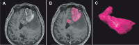
Quantification of the preoperative tumour volume in malignant gliomas. (A) Axial T1-weighted MRI scan of a patient with a left frontal glioblastoma. (B) The contrast-enhancing part of each section is outlined and subsequently fused to generate (C) a 3-dimensional segment which can be quantified volumetrically.

Keywords: glioma,  MRI,  Tumor
7. Proescholdt M; Doenitz C; Brawanski A
Surgery of Malignant Gliomas Using Modern Technology
Glioma - Surgery
European Association of NeuroOncology Magazine 2013; 3 (1): 11-14
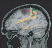
Fusion of [F-18]fluoroethyltyrosine: PET (red), functional MRI (green), and DTI tractography (yellow) in a patient with a left frontal anaplastic astrocytoma. Note the close vicinity of eloquent cortical and subcortical structures to the tumour borders.

Keywords: glioma,  MRI,  PET
8. Bello L; Riva M; Casaceli G; Fava E
Technological Advances in Glioma Surgery
DTI-FT
European Association of NeuroOncology Magazine 2011; 1 (1): 13-19
Example of integration of intraoperative neurophysiology with advanced MR imaging (DTI-FT) in a case of a left rolandic tumour. In this case, at the beginning of the resection the surgeon should integrate information coming from DES cortical mapping (a) (left and middle panel showing hand motor responses from the hand obtained by train of 5 techniques [left panel]) and 60 Hz probes [middle panel]), to those obtained by DTI-FT. The right panel shows the DTI-FT reconstruction for CST (in white) superimposed onto post-contrast T1-weighted images. The green cross indicates the position of the cortex where DES found hand motor responses. The site corresponded to the CST as indicated by DTI-FT images. The same type of integration is shown during resection at the subcortical level (b). The green cross indicates the site where DES (with both train of 5 techniques and 60 Hz) located hand responses from the subcortical of the CST, which corresponded to the same position in the DTI-FT images.

Keywords: CST,  DTI-FT,  MRI
9. Bello L; Riva M; Casaceli G; Fava E
Technological Advances in Glioma Surgery
Operating Theatre
European Association of NeuroOncology Magazine 2011; 1 (1): 13-19
Example of integration in the operating theatre of various types of imaging in a case of left temporo-insular low-grade glioma: (a) volumetric FLAIR; (b) DTI FT reconstruction of IFO (white) superimposed onto a postcontrast T1-weighted MR image; (c) spots of activation for denomination (in white) obtained with fMRI, superimposed onto FLAIR images; (d) axial and (e) coronal post-contrast-T1 weighted images, and (f) spots of activation for verbal generation (in white) obtained with fMRI, superimposed onto FLAIR images. All these images were loaded into the neuronavigation system, co-registered, and fused together to be available during surgery. Surgery was performed in awake anaesthesia and the patient was continuously submitted to object-naming tests by the neuropsychologist during resection. The green cross indicates a subcortical site where semantic paraphrasia was induced by DES, at this site corresponded to the IFO as indicated in (b) and to the medial deep border of the tumour, as shown by FLAIR images (a).

Keywords: FLAIR,  MRI

 

 Seite 1 von 1 
 
copyright © 2000–2026 Krause & Pachernegg GmbH | Sitemap | Datenschutz | Impressum
 
Werbung