| Halestrap AP |
|---|
The Mitochondrial Permeability Transition - A Pore Way for the Heart to Die
Journal of Clinical and Basic Cardiology 2002; 5 (1): 29-41
PDF Summary Figures
| Figure |
|---|
| |
|
|
Mitochondrien - Permeabilität
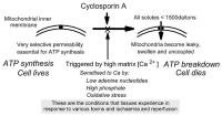
Figure 1: The essential features of the permeability transition and its potential role in necrotic cell death
Keywords: Membran,
Membrane,
Mitochondria,
Mitochondrium,
permeability,
Permeabilität,
Schema
|
| |
| |
|
|
Mitochondrien - Apoptose
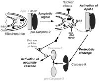
Figure 2: The release of mitochondrial proteins plays a central role in the initiation of apoptosis. An apoptotic signal leads to the release of pro-apoptotic proteins located within the intermembrane space between the inner and outer mitochondrial membranes (IMM and OMM) into the cytosol. The pathway for release may be OMM rupture following opening of the MPTP and mitochondrial swelling, or through recruitment of specific pore-forming proteins to the outer
membrane such as Bax and t-Bid. Release is inhibited by Bcl-2. The released cytochrome binds to Apaf-1 that proteolytically activates caspase 9 which in turn activates caspase 3 and so initiates
the apoptotic cascade. Caspases are inhibited by endogenous inhibitors which are themselves inactivated by Smac (or DIABLO) that is also released along with apoptosis inducing factor (AIF).
This migrates to the nucleus where it causes chromatin condensation and DNA fragmentation. Further details are given in the text.
Keywords: Apoptose,
apoptosis,
Mitochondrion,
Mitochondrium,
Schema
|
| |
| |
|
|
Mitochondrien - Permeabilität
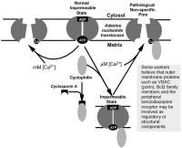
Figure 3: The proposed mechanism of the mitochondrial permeability transition pore. The probable sites of action of known effectors of pore opening are shown in Table 1.
Keywords: Mitochondrion,
Mitochondrium,
permeability,
Permeabilität,
Pore,
Pore,
Schema
|
| |
| |
|
|
Mitochondrien - Permeabilität
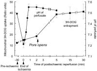
Figure 4: Time dependence of MPTP opening during reperfusion. The opening of the MPTP was detected using the DOG preloading technique. Hearts were subjected to 30 min global ischaemia before reperfusion for the time shown. Data are taken from [77]. Parallel data for the pH of the perfusate are taken from [97].
Keywords: Diagramm,
ischaemia,
Ischämie,
Mitochondrion,
Mitochondrium,
permeability,
Permeabilität,
Pore,
Pore,
reperfusion,
reperfusion
|
| |
| |
|
|
Mitochondrien - Permeabilität
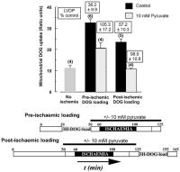
Figure 5: Pyruvate protects the heart against reperfusion injury through inhibiting MPTP opening and enhancing subsequent pore closure. Pore opening upon reperfusion was determined using the entrapment of pre-loaded 3H-DOG, whilst subsequent closure of the MPTP was detected by measuring the entrapment of 3H-DOG loaded after maximal recovery of the heart (post-loading). These protocols are illustrated in the Figure and further details are given in the text and reference [97] from where the data are taken.
Keywords: Diagramm,
ischaemia,
Ischämie,
Mitochondrion,
Mitochondrium,
Pore,
Pore,
Pyruvat,
Pyruvate
|
| |
| |
|
|
Mitochondrien - Apoptose
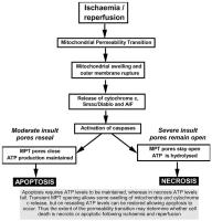
Figure 6: A scheme to illustrate how the extent and permanence of MPTP opening may determine whether heart cells die by apoptosis or necrosis.
Keywords: Apoptose,
apoptosis,
ischaemia,
Ischämie,
Mitochondrion,
Mitochondrium,
necrosis,
Nekrose,
reperfusion,
reperfusion,
Schema
|
| |
| |
|
|

