| Zijdewind J et al. | ||||||||||||||||
|---|---|---|---|---|---|---|---|---|---|---|---|---|---|---|---|---|
|
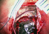
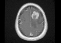
Case Report: Primary Leptomeningeal Melanoma in a Patient with Neurocutaneous Melanosis
European Association of NeuroOncology Magazine 2013; 3 (3): 141-142 PDF Figures
|
||||||||||||||||

Verlag für Medizin und Wirtschaft |
|
||||||||
|
Figures and Graphics
|
|||||||||
| copyright © 2000–2025 Krause & Pachernegg GmbH | Sitemap | Datenschutz | Impressum | |||||||||

