Zijdewind J, Wesseling P, de Witt Hamer PC, Reijneveld J Case Report: Primary Leptomeningeal Melanoma in a Patient with Neurocutaneous Melanosis European Association of NeuroOncology Magazine 2013; 3 (3): 141-142 PDF Overview
| ||||
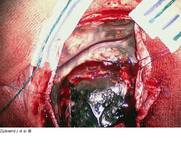
Figure/Graphic 1: Leptomeningeal Tumour Intraoperative photograph after opening of the dura, showing a black leptomeningeal tumour in the left frontal region. Of note is the spotty black appearance of the arachnoid, which is not continuous with the tumour, consistent with neurocutaneous melanosis. |

Figure/Graphic 1: Leptomeningeal Tumour
Intraoperative photograph after opening of the dura, showing a black leptomeningeal tumour in the left frontal region. Of note is the spotty black appearance of the arachnoid, which is not continuous with the tumour, consistent with neurocutaneous melanosis. |


