Krause und Pachernegg
Verlag für Medizin und Wirtschaft
Artikel   Bilder   Volltext

Mobile Version
A-  |   A  |   A+
Werbung
 
Abbildungen und Graphiken
Baimbetov AK et al.  
Immediate Results of Pulmonary Veins Entries Cryoblation of Patients with Atrial Fibrillation // Direkte Ergebnisse der Kryo-Ablation der PV-Ostien bei Patienten mit Vorhofflimmern

Zeitschrift für Gefäßmedizin 2018; 15 (3): 11-16

Volltext (PDF)    Summary    Abbildungen   


Abbildung
 
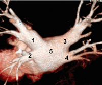
Multispiral CT
Abbildung 1: Multispiral computed tomography of left atrium and pulmonary veins (back view). 1: LUPV, 2: LLPV, 3: RUPV, 4: RLPV, 5: left atrium back wall.


Keywords: atrial fibrillationcryoablationGefäßmedizinleft atriumMultispiral CTpulmonary veins
 
 
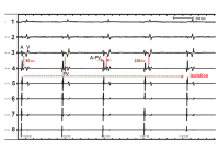
ECG and endograms
Abbildung 2: Record of ECG and endograms during pulmonary veins entries cryoblation. 1: surface ECG (2nd derivation); Endograms: 2–4: mapping “achieve” catheter (LS 2–8), 5–8: diagnostic probe within coronary sinus; Potentials: А) for atriums, V) for ventricles, PV) for pulmonary veins; Gradual blocking of PV potentials and their total disappearance upon reaching PV isolation from LA is displayed.


Keywords: atrial fibrillationcryoablationECGendogramGefäßmedizin
 
 
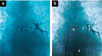
Cryoablation of LUPV
Abbildung 3a-b: Cryoablation of LUPV. Full obstruction of target vein. 1: Cryoballoon in inflated state at LUPV entry, 2: delivery catheter to LA, 3: 10-polar probe within coronary sinus, 4–8: polar “achieve” catheter in LUPV, 5: contrast stagnation confirms full obstruction of ablating PV


Keywords: atrial fibrillationcryoablationcryoballoonGefäßmedizinLUPV
 
 
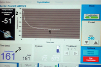
Cryoconsole display
Abbildung 4: Cryoconsole display during ablation. 1: the curve shows the reached temperature at the point of contact of cryoballon with tissue of LA and PVE, 2: temperature indicator, 3: cryotherapy time indicator


Keywords: atrial fibrillationcryoablationcryoconsole displayGefäßmedizin


copyright © 2000–2025 Krause & Pachernegg GmbH | Sitemap | Datenschutz | Impressum
 
Werbung