Krause und Pachernegg
Verlag für Medizin und Wirtschaft
Artikel   Bilder   Volltext

Mobile Version
A-  |   A  |   A+
Werbung
 
Abbildungen und Graphiken
Kasseroller R  
LVF-Lymphödemklassifikation des inguinalen und axillären Tributargebietes

Zeitschrift für Gefäßmedizin 2005; 2 (4): 4-8

Volltext (PDF)    Summary    Abbildungen   


Abbildung
 
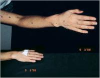
Hautfaltenmessung
Abbildung 1: Hautfaltenmeßpunkte Normalwert < 2 mm


Keywords: FotoGefäßmedizinHautfaltenmesspunktLymphologie
 
 
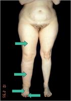
Hautfaltenmessung
Abbildung 2: Meßpunkte an den unteren Extremitäten


Keywords: FotoGefäßmedizinHautfaltenmesspunktLymphologie
 
 
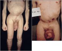
Hautfaltenmessung
Abbildung 3: Hautfaltenmeßpunkte am Thorax. Stern 1: Schnittpunkt Axillarlinie – Mitte-Sternum-Linie; Stern 2: Schnittpunkt Medioklavikularlinie – Mitte-Sternum-Linie; Stern 3: Halbierende auf Medioklavikularlinie – Clavicula-Mitte-Sternum-Linie


Keywords: GefäßmedizinHautfaltenmesspunktLymphologieSchemaThorax
 
 
Fibrotisches Lymphödem
Abbildung 4: ... er ist völlig gesund ...


Keywords: FibroseFotoGefäßmedizinLymphologieLymphödem


copyright © 2000–2025 Krause & Pachernegg GmbH | Sitemap | Datenschutz | Impressum
 
Werbung