Trophoblast turnover
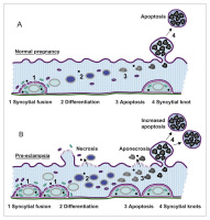
Abbildung 1: Schematic representation of trophoblast turnover. (A) Normal pregnancy: The final event of cytotrophoblast differentiation, syncytial fusion, results in incorporation of fresh organelles and other cellular
material into the syncytium (1). Within the multinucleated syncytiotrophoblast
differentiation (2) and subsequently late apoptosis (3) take place. Finally, the late apoptotic material is packed into protrusions of the apical plasma membrane, syncytial knots. These knots are released into the maternal circulation as tightly sealed corpuscles (4). (B) Pre-eclampsia: Enhanced proliferation and syncytial fusion (1) may overwhelm the
capacity of the syncytiotrophoblast in terms of differentiation (2) and
apoptotic release (3). This may result in a necrotic breakdown of specific
sites of the syncytiotrophoblast. If at these sites apoptosis has not yet started, pure necrosis can be observed; if however apoptosis has already lead to first cleavage of proteins, apoptotic material will be necrotically released (aponecrosis). At the same time the syncytiotrophoblast tries to counter balance for the increased input by increasing the release of apoptotic syncytial knots (4). Modified from [15].
Keywords:
pregnancy,
Schema,
scheme,
Schwangerschaft,
trophoblast


