Huppertz B Placental Villous Trophoblast: the Altered Balance Between Proliferation and Apoptosis Triggers Pre-eclampsia Journal für Reproduktionsmedizin und Endokrinologie - Journal of Reproductive Medicine and Endocrinology 2006; 3 (2): 103-108 Volltext (PDF) Summary Übersicht
| ||||
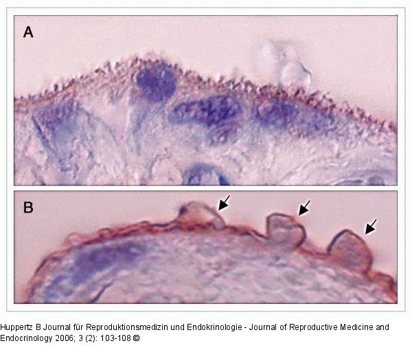
Abbildung 2: Term placentas Representative images of chorionic villi from term placentas derived from a normal pregnancy (A) and a pregnancy complicated by late-onset pre-eclampsia (B). In both images staining was performed using a primary antibody against placental-protein 13 (PP13), which is present in the apical membrane of the syncytiotrophoblast. In (A) the normal villous brush border can be seen, which is stained for PP13. In (B) the alterations of the brush border membrane are obvious. Protrusions on the cellular level can be seen (arrows) that may be released into the maternal circulation. Magnification ×900. |

Abbildung 2: Term placentas
Representative images of chorionic villi from term placentas derived from a normal pregnancy (A) and a pregnancy complicated by late-onset pre-eclampsia (B). In both images staining was performed using a primary antibody against placental-protein 13 (PP13), which is present in the apical membrane of the syncytiotrophoblast. In (A) the normal villous brush border can be seen, which is stained for PP13. In (B) the alterations of the brush border membrane are obvious. Protrusions on the cellular level can be seen (arrows) that may be released into the maternal circulation. Magnification ×900. |


