| Zeller T et al. |
|---|
Belastungs-Duplexsonographie zur Verlaufskontrolle nach Arteria thoracica interna-Bypass auf den Ramus interventricularis anterior in MIDCAB-Technik: Beschreibung der Methodik und eigene Ergebnisse
Zeitschrift für Gefäßmedizin 2006; 3 (4): 4-10
Volltext (PDF) Summary Abbildungen
| Abbildung |
|---|
| |
|
|
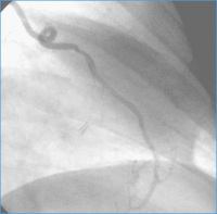
ATI-Bypass
Abbildung 1: Beispiel eines ATI-Bypasses auf den Ramus interventricularis anterior 8 Jahre nach Implantation in MIDCAB-Technik.
Keywords: Angiographie,
Arteria thoracica interna,
Bildgebende Diagnostik,
Bypass,
Gefäßmedizin
|
| |
| |
|
|
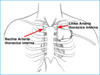
Arteria thoracica interna
Abbildung 2: Topographie A. throracica interna beidseits. Lage des Ultraschallkopfes
Keywords: Arteria thoracica interna,
Bildgebende Diagnostik,
Gefäßmedizin,
Schema
|
| |
| |
|
|
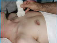
Arteria thoracica interna
Abbildung 3a: Schallkopfposition bei Untersuchung der A. thoracica interna
Keywords: Arteria thoracica interna,
Bildgebende Diagnostik,
Foto,
Gefäßmedizin
|
| |
| |
|
|
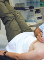
Arteria thoracica interna
Abbildung 3b: Position der Beine bei Azoulay-Test
Keywords: Arteria thoracica interna,
Azoulay-Test,
Bildgebende Diagnostik,
Foto,
Gefäßmedizin
|
| |
| |
|
|
Arteria thoracica interna
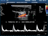
Abbildung 4: Triphasisches normales Dopplerprofil: Linke A. thoracica interna vor Bypassanastomosierung.
Keywords: Arteria thoracica interna,
Bildgebende Diagnostik,
Duplexsonographie,
Gefäßmedizin
|
| |
| |
|
|
Arteria thoracica interna
Abbildung 4b: Mono- bis biphasisches Dopplerspektrum mit kurzem retrogradem Fluß enddiastolisch der A. thoracica interna nach MIDCAB-Operation.
Keywords: Arteria thoracica interna,
Bildgebende Diagnostik,
Duplexsonographie,
Gefäßmedizin
|
| |
| |
|
|
Bypassverschluß
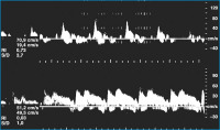
Abbildung 4b: Dopplerprofil bei Bypassverschluß und nach Revision.
Keywords: Bildgebende Diagnostik,
Bypassverschluss,
Duplexsonographie,
Gefäßmedizin
|
| |
| |
|
|
Volumenbelastung
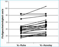
Abbildung 5: Patienten ohne Bypass-Stenose: Zunahme der distolischen Spitzenflußgeschwindigkeiten
(Vd) unter Volumenbelastung.
Keywords: Azoulay-Test,
Bildgebende Diagnostik,
Duplexsonographie,
Gefäßmedizin,
Volumenbelastung
|
| |
| |
|
|
Volumenbelastung
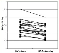
Abbildung 6: Patienten ohne Bypass-Stenose: Abnahme des systolisch/diastolischen
Flußquotienten in Ruhe unter Volumenbelastung.
Keywords: Azoulay-Test,
Bildgebende Diagnostik,
Duplexsonographie,
Gefäßmedizin,
Volumenbelastung
|
| |
| |
|
|
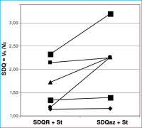
Volumenbelastung
Abbildung 7: Patienten mit Bypass-Stenosen: Anstieg des systolisch/diastolischen Flußquotienten unter Volumenbelastung (SDQ az + St) im Vergleich zum Ruhewert (SDQ R + St).
Keywords: Azoulay-Test,
Bildgebende Diagnostik,
Duplexsonographie,
Gefäßmedizin,
Volumenbelastung
|
| |
| |
|
|
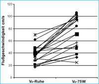
Fahrradergometrie
Abbildung 8: Patienten ohne Bypass-Stenose: Anstieg der diastolischen Spitzenflußgeschwindigkeit (Vd)
unter Fahrradergometrie bei 75 W.
Keywords: Bildgebende Diagnostik,
Bypassgesund,
Fahrradergometrie,
Gefäßmedizin
|
| |
| |
|
|
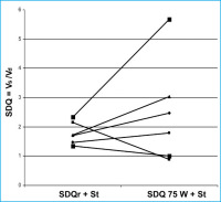
Fahrradergometrie
Abbildung 9: Patienten mit Bypass-Stenosen: Anstieg des systolisch/diastolischen Flußquotienten unter Fahrradergometerbelastung bei 75 W (SDQ 75 W + St) im Vergleich zum Ruhewert (SDQ R + St).
Keywords: Bildgebende Diagnostik,
Bypass,
Fahrradergometrie,
Gefäßmedizin
|
| |
| |
|
|
Volumenbelastung - Ergometerbelastung
Abbildung 10: Patienten ohne Bypass-Stenose: Vergleich Volumenbelastung und
Ergometerbelastung bei 75 W: Anstieg der diastolischen Spitzenflußgeschwindigkeit
(Vd).
Keywords: Bildgebende Diagnostik,
Bypassgesund,
Fahrradergometrie,
Gefäßmedizin,
Volumenbelastung
|
| |
| |
|
|

