Krause und Pachernegg
Verlag für Medizin und Wirtschaft
Artikel   Bilder   Volltext

Mobile Version
A-  |   A  |   A+
Werbung
 
Abbildungen und Graphiken
Jacobeit JW, Kliesch S  
DFP/CME: Diagnostik und Therapie der Gynäkomastie. Nicht jede Brustdrüsenvergrößerung des Mannes ist eine Gynäkomastie, aber jede Gynäkomastie ist abklärungsbedürftig.

Journal für Reproduktionsmedizin und Endokrinologie - Journal of Reproductive Medicine and Endocrinology 2009; 6 (2): 63-67

Volltext (PDF)    Summary    Praxisrelevanz    Abbildungen   


Abbildung
 
Gynäkomastie
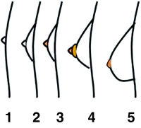
Abbildung 1: Tanner-Stadien (aus: Wikipedia. Die freie Enzyklopädie, GNU-Lizenz für freie Dokumentation).


Keywords: GynäkomastieTanner-Stadien
 
 
Gynäkomastie
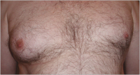
Abbildung 2: 47-jähriger Patient, Anisomastie bei Makromastie Stadium B 5 (BI-RADS 3) infolge unbehandeltem Kallmann-Syndrom.


Keywords: FotoGynäkomastie
 
 
Gynäkomastie
Abbildung 3: 37-jähriger Patient mit neu aufgetretener schmerzhafter einseitiger Gynäkomastie rechts (BI-RADS 3) mit OP-Indikation.


Keywords: FotoGynäkomastie
 
 
Gynäkomastie
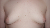
Abbildung 4: 21-jähriger Patient mit komplett fibrosierter Pubertätsgynäkomastie ohne Regredienz (BI-RADS 1) – Indikation für beidseitige Mastektomie und Fettabsaugung


Keywords: FotoGynäkomastie
 
 
Gynäkomastie
Abbildung 5: 34-jähriger Patient, ausgeprägte schmerzhafte Brustschwellung bds. Diagnose: akute TRAK-positive ausgeprägte Hyperthyreose M. Basedow (BI-RADS 2).


Keywords: FotoGynäkomastie


copyright © 2000–2025 Krause & Pachernegg GmbH | Sitemap | Datenschutz | Impressum
 
Werbung