Krause und Pachernegg
Verlag für Medizin und Wirtschaft
Artikel   Bilder   Volltext

Mobile Version
A-  |   A  |   A+
Werbung
 
Abbildungen und Graphiken
Ure C, Döller W  
Extremitätenlymphödem - Diagnosesicherung durch einen diagnostischen Algorithmus

Zeitschrift für Gefäßmedizin 2011; 8 (2): 5-8

Volltext (PDF)    Summary    Praxisrelevanz    Abbildungen   


Abbildung
 
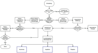
lymphologische Basisdiagnostik
Abbildung 1: Workflow der lymphologischen Basisdiagnostik. Quelle: Zentrum für Lymphologie LKH-Wolfsberg.


Keywords: Gefäßmedizinlymphologische BasisdiagnostikSchemaWorkflow
 
 
lymhpologische Zusatzdiagnostik
Abbildung 2: Workflow der lymphologischen Zusatzdiagnostik. Quelle: Zentrum für Lymphologie LKH-Wolfsberg.


Keywords: Gefäßmedizinlymphologische ZusatzdiagnostikSchemaWorkflow
 
 
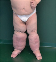
Chronisches Beinlymphödem
Abbildung 3: Chronisches Beinlymphödem im Stadium III vor komplexer physikalischer Entstauungstherapie KPE (Datum: 04.08.2010). Quelle: Zentrum für Lymphologie LKH-Wolfsberg.


Keywords: Chronisches BeinlymphödemEntstauungstherapieGefäßmedizinKPEStadium III
 
 
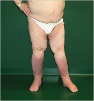
Chronisches Beinlymphödem nach 3 Wochen
Abbildung 4: Nach 3-wöchiger stationärer KPE (Datum: 13.09.2010). Quelle: Zentrum für Lymphologie LKH-Wolfsberg.


Keywords: Chronisches BeinlymphödemEntstauungstherapieFotoGefäßmedizinstationäre KPE


copyright © 2000–2025 Krause & Pachernegg GmbH | Sitemap | Datenschutz | Impressum
 
Werbung