Schellong SM Die Ultraschalluntersuchung der Beinvenen bei Verdacht auf Venenthrombose - eine Übersicht Zeitschrift für Gefäßmedizin 2009; 6 (3): 5-10 Volltext (PDF) Summary Übersicht
| ||||||||
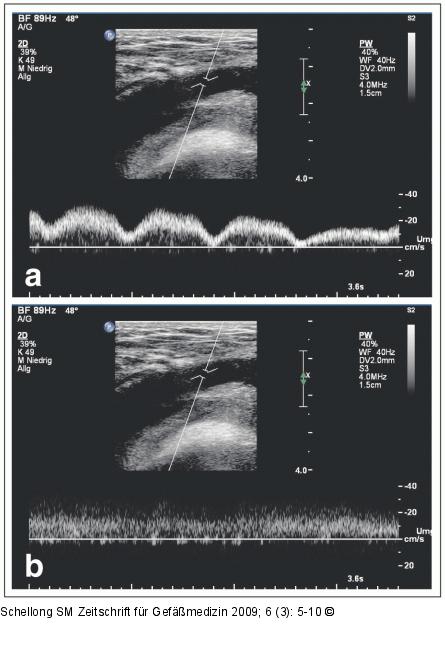
Abbildung 4a-b: Vena femoralis communis PW-Dopplersignal aus der Vena femoralis communis. a: Normales Strömungsprofil mit Atem- und Pulsmodulation. b: Bandförmiges Signal als Hinweis auf ein proximal gelegenes Strömungshindernis. |

Abbildung 4a-b: Vena femoralis communis
PW-Dopplersignal aus der Vena femoralis communis. a: Normales Strömungsprofil mit Atem- und Pulsmodulation. b: Bandförmiges Signal als Hinweis auf ein proximal gelegenes Strömungshindernis. |


